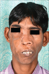
Giant odontogenic fibroma of maxilla
- PMID: 25593878
- PMCID: PMC4293849
- DOI: 10.4103/2231-0746.147148
Giant odontogenic fibroma of maxilla
Abstract
Odontogenic fibroma is a benign ectomesenchymal tumor classified as central and peripheral on the basis of its location and as an epithelium rich or epithelium poor based on its histological features. Radiological findings consist of radiolucent areas with well-defined bony margins. The lesion is detected early because of its location and usually treated with surgical excision and curettage. We present a case of giant odontogenic fibroma of right maxilla presenting as gross facial deformity and posing a dual challenge of excising the tumor mass and reconstructing the ensuing defect.
Keywords: Giant; maxilla; odontogenic fibroma; reconstruction.
Conflict of interest statement
Figures


References
-
- Wesley RK, Wysocki GP, Mintz SM. The central odontogenic fibroma. Clinical and morphologic studies. Oral Surg Oral Med Oral Pathol. 1975;40:235–45. - PubMed
-
- Barnes L, Eveson JW, Reichart P, Sidransky D. Pathology and Genetics of Head and Neck Tumors. Lyon: IARC Press; 2005. World Health Organization Classification of Tumors; p. 317.
-
- Raubenheimer EJ, Noffke CE. Central odontogenic fibroma-like tumors, hypodontia, and enamel dysplasia: Review of the literature and report of a case. Oral Surg Oral Med Oral Pathol Oral Radiol Endod. 2002;94:74–7. - PubMed
-
- Allen CM, Hammond HL, Stimson PG. Central odontogenic fibroma, WHO type. A report of three cases with an unusual associated giant cell reaction. Oral Surg Oral Med Oral Pathol. 1992;73:62–6. - PubMed
-
- Daniels JS. Central odontogenic fibroma of mandible: A case report and review of the literature. Oral Surg Oral Med Oral Pathol Oral Radiol Endod. 2004;98:295–300. - PubMed
Publication types
LinkOut - more resources
Full Text Sources
Other Literature Sources
