Review
. 2014 Nov 18:1:44.
doi: 10.3389/fmed.2014.00044. eCollection 2014.

Pretargeted imaging and radioimmunotherapy of cancer using antibodies and bioorthogonal chemistry

Affiliations
Review

Pretargeted imaging and radioimmunotherapy of cancer using antibodies and bioorthogonal chemistry

Floor C J van de Watering et al. Front Med (Lausanne). .

Abstract

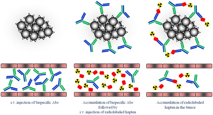
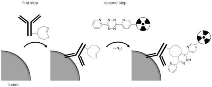
Selective delivery of radionuclides to tumors may be accomplished using a two-step approach, in which in the first step the tumor is pretargeted with an unlabeled antibody construct and in the second step the tumor is targeted with a radiolabeled small molecule. This results in a more rapid clearance of the radioactivity from normal tissues due to the fast pharmacokinetics of the small molecule as compared to antibodies. In the last decade, several pretargeting approaches have been tested, which have shown improved tumor-to-background ratios and thus improved imaging and therapy as compared to directly labeled antibodies. In this review, we will discuss the strategies and applications in (pre-)clinical studies of pretargeting concepts based on the use of bispecific antibodies, which are capable of binding to both a target antigen and a radiolabeled peptide. So far, three generations of the bispecific antibody-based pretargeting approach have been studied. The first clinical studies have shown the feasibility and potential for these pretargeting systems to detect and treat tumor lesions. However, to fully integrate the pretargeting approach in clinic, further research should focus on the best regime and pretargeting protocol. Additionally, recent developments in the use of bioorthogonal chemistry for pretargeting of tumors suggest that this chemical pretargeting approach is an attractive alternative strategy for the detection and treatment of tumor lesions.

Keywords: bispecific antibodies; pretargeting; radioimmunodetection; radioimmunotherapy; tumor-associated antigen.


Figures

Figure 1
Figure 1
Schematic overview of the pretargeting strategy. First, tumors are pretargeted with bispecific antibodies (bsAb). Secondly, small radiolabeled hapten peptide is i.v. injected and binds to the pretargeted bsAb at the tumor cells.
Figure 2
Figure 2
Biodistribution of 111In-diDTPA (A) and 111In-DTPA (B) at 1?72 h after injection of the radioactivity in nude mice with s.c. NU-12 tumors. Mice received 100 pmol of G250 × DTIn1 bsAb i.v. and 3 days later 7 pmol of 111In-DTPA or 111In-diDTPA. As a reference a separate set of nude mice with s.c. NU-12 xenografts received the directly labeled antibody 111In-DTPA-G250 (C). This research was originally published in Cancer Research by Boerman et al. (16).
Figure 3
Figure 3
Schematic representation of how a trivalent bispecific antibody is formed by the DNL method. Component A links a cysteine-modified dimerization and docking domain (DDD) to a Fab of an anti-tumor antibody, which results in spontaneous formation of component A2. Component B links an anchoring domain to a Fab of an anti-hapten antibody. The AD is modified with cysteine on each end. AD will naturally “dock” with the DDD when components A and B are mixed, which brings the two molecules together in a well-defined orientation and also results in disulfide bonds across the two proteins. This research was originally published in Goldenberg et al. (42).
Figure 4
Figure 4
PET/CT images of a BALB/c nude mouse with a s.c. LS174T tumor on the right hind leg (arrow) and an inflammation in the left thigh muscle (arrowhead), which received 18F-FDG and, 1 day later, 6.0 nmol TF2 and 68Ga-IMP288 (0.25 nmol) with a 16-h interval. The animal was imaged 1 h after 18F-FDG and 68Ga-IMP288 injections. The panel shows the three-dimensional volume rendering the pretargeted immuno-PET scan (A) and the FDG-PET scan (B), and the transverse sections of the tumor region of the pretargeted immuno-PET scan (C) and the FDG-PET scan (D). This research was originally published in Schoffelen et al. (48, 50).
Figure 5
Figure 5
The SPECT/CT image (A), acquired 24 h after injection of 111In-IMP288 (185 MBq, 25 μg), pretargeted with 75 mg TF2 (1-day interval), in a 38-year-old patient (cohort 4), shows very clear tumor targeting of an axillary lymph-node metastasis, with very low concentrations of radioactivity in normal tissues. Corresponding contrast-enhanced CT scan and a fused FDG-PET/CT scan are shown [(B,C), respectively]. The primary colon tumor (50 cm ab ano) also shows highly specific tumor targeting in the SPECT image (D), confirmed by the CT scan and FDG-PET/CT [(E,F), respectively], with non-specific FDG uptake in the ascending colon. This research was originally published in Schoffelen et al. (52).
Figure 6
Figure 6
Schematic overview of tumor pretargeting by using the inverse-electron-demand Diels–Alder reaction. This research was originally published in Rossin et al. (60).
Figure 7
Figure 7
SPECT/CT imaging of mice bearing colon carcinoma xenografts: posterior projections of mice preinjected with (A) CC49-TCO followed 1 day later by 111In-DOTA-tetrazine (1:25, 42 MBq), (B) CC49 followed 1 day later by 111In-DOTA-tetrazine (1:25, 20 MBq), (C) irrelevant Ab (Rtx-TCO; 100 μg) followed 1 day later by 111In-DOTA-tetrazine (1:25, 50 MBq), (D–F) single transverse slices (2 mm) passing through the tumors in (A–C). This research was originally published in Rossin et al. (60).
Figure 8
Figure 8
PET images of 64Cu-Tz-Bn-NOTA/A33-TCO pretargeting strategy, 64Cu-NOTA-A33 and 89Zr-DFO-A33.Transverse (top) and coronal (bottom) planar images intersect the center of the tumors. This research was originally published in Zeglis et al. (63).

References

    1. Fleuren EDG, Versleijen-Jonkers YMH, Heskamp S, van Herpen CML, Oyen WJG, van der Graaf WTA, et al. Theranostic applications of antibodies in oncology. Mol Oncol (2014) 8(4):799–812 10.1016/j.molonc.2014.03.010 - DOI - PMC - PubMed
    1. Heskamp S, van Laarhoven HW, Oyen WJ, van der Graaf WT, Boerman OC. Tumor-receptor imaging in breast cancer: a tool for patient selection and response monitoring. Curr Mol Med (2013) 13(10):1506–22. 10.2174/1566524013666131111120638 - DOI - PubMed
    1. Goldenberg DM, Sharkey RM. Using antibodies to target cancer therapeutics. Expert Opin Biol Ther (2012) 12(9):1173–90. 10.1517/14712598.2012.693472 - DOI - PubMed
    1. Boerman OC, van Schaijk FG, Oyen WJG, Corstens FHM. Pretargeted radioimmunotherapy of cancer: progress step by step. J Nucl Med (2003) 44(3):400–11. - PubMed
    1. Kong F-L, Ford RJ, Yang DJ. Managing lymphoma with non-FDG radiotracers: current clinical and preclinical applications. Biomed Res Int (2013) 2013:12. 10.1155/2013/626910 - DOI - PMC - PubMed