

Review
doi: 10.3389/fmed.2014.00048.
eCollection 2014.
Placental amniotic epithelial cells and their therapeutic potential in liver diseases
Affiliations
- PMID: 25593921
- PMCID: PMC4291892
- DOI: 10.3389/fmed.2014.00048
Item in Clipboard
Review
Placental amniotic epithelial cells and their therapeutic potential in liver diseases
Front Med (Lausanne).
.
Abstract
As a unique source of stem cells, there is a growing interest in amniotic epithelial (AE) cells. Placenta is readily available; in fact, it is often discarded following delivery. As such, it is without the ethical concerns of embryonic stem cells. Further advantages to AE include that AE cells do not demonstrate tumorigenicity upon transplantation, and are gifted with immunomodulatory and anti-inflammatory properties. Thus, AE cells have exceptional features for use as cell-based therapies for liver disease.
Keywords: liver diseases; placenta; placental amniotic epithelial cells; stem cells; therapy.
Figures
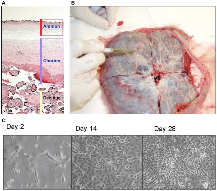
(A) Human placenta layers: amnion, chorion, and decidua. Amniotic layer is composed of a single-celled epithelial layer and a deeper mesodermal layer. Chorionic layer is composed of a mesodermal layer and a trophoblast layer. (B) Isolation of amnion membrane from placenta. The maternal side of placenta is placed face down and a shallow X-shaped incision is made through the center of the placenta. The thin, nearly transparent amnion membrane is then peeled starting at the center of the cut and progressing outward. (C) Morphology of amniotic epithelial cells in culture (×40).
Similar articles
-
Therapeutic potential of placenta-derived stem cells for liver diseases: current status and perspectives.J Obstet Gynaecol Res. 2014 Feb;40(2):360-8. doi: 10.1111/jog.12213. Epub 2013 Nov 18. J Obstet Gynaecol Res. 2014. PMID: 24245961 Review.
-
Human placental stem cells: biomedical potential and clinical relevance.J Stem Cells. 2011;6(2):75-92. J Stem Cells. 2011. PMID: 22997848 Review.
-
Human amniotic epithelial stem cells: Hepatic differentiation and regenerative properties in liver disease treatment.Placenta. 2023 Mar 24;134:39-47. doi: 10.1016/j.placenta.2023.02.013. Epub 2023 Feb 28. Placenta. 2023. PMID: 36870301 Review.
-
A Rational Strategy for the Use of Amniotic Epithelial Stem Cell Therapy for Liver Diseases.Stem Cells Transl Med. 2016 Apr;5(4):405-9. doi: 10.5966/sctm.2015-0304. Epub 2016 Mar 3. Stem Cells Transl Med. 2016. PMID: 26941361 Free PMC article. Review.
-
Amniotic fluid and placental membranes: unexpected sources of highly multipotent cells.Semin Reprod Med. 2013 Jan;31(1):62-8. doi: 10.1055/s-0032-1331799. Epub 2013 Jan 17. Semin Reprod Med. 2013. PMID: 23329638 Review.
Cited by
-
Primary Broiler Hepatocytes for Establishment of a Steatosis Model.Vet Sci. 2022 Jun 24;9(7):316. doi: 10.3390/vetsci9070316. Vet Sci. 2022. PMID: 35878333 Free PMC article.
-
Therapeutic Effects of Human Amniotic Epithelial Stem Cells in a Transgenic Mouse Model of Alzheimer's Disease.Int J Mol Sci. 2020 Apr 10;21(7):2658. doi: 10.3390/ijms21072658. Int J Mol Sci. 2020. PMID: 32290355 Free PMC article.
-
Hepatocyte transplantation and advancements in alternative cell sources for liver-based regenerative medicine.J Mol Med (Berl). 2018 Jun;96(6):469-481. doi: 10.1007/s00109-018-1638-5. Epub 2018 Apr 24. J Mol Med (Berl). 2018. PMID: 29691598 Free PMC article. Review.
-
Pregnancy state before the onset of labor: a holistic mechanical perspective.Biomech Model Mechanobiol. 2024 Oct;23(5):1531-1550. doi: 10.1007/s10237-024-01853-3. Epub 2024 May 17. Biomech Model Mechanobiol. 2024. PMID: 38758337 Free PMC article.
-
Cell-based approaches for augmentation of tendon repair.Tech Shoulder Elb Surg. 2017 Sep;18(3):e6-e14. doi: 10.1097/BTE.0000000000000132. Epub 2017 Sep 1. Tech Shoulder Elb Surg. 2017. PMID: 29276433 Free PMC article.
References
Publication types
LinkOut - more resources
Full Text Sources
Other Literature Sources
Research Materials