Review
. 2014 Sep 25:1:38.
doi: 10.3389/fsurg.2014.00038. eCollection 2014.

Biology of infantile hemangioma

Affiliations
Review

Biology of infantile hemangioma

Tinte Itinteang et al. Front Surg. .

Abstract

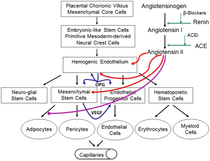
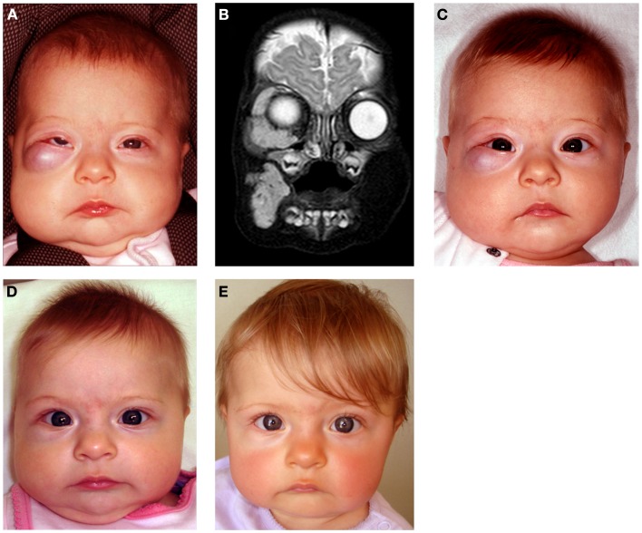
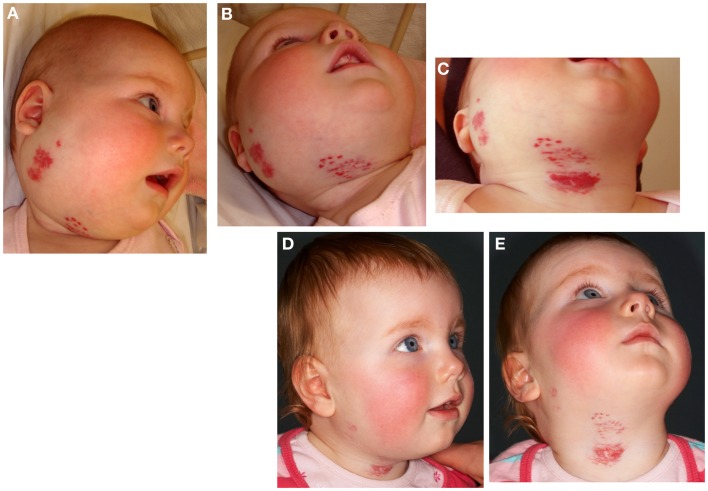
Infantile hemangioma (IH), the most common tumor of infancy, is characterized by an initial proliferation during infancy followed by spontaneous involution over the next 5-10 years, often leaving a fibro-fatty residuum. IH is traditionally considered a tumor of the microvasculature. However, recent data show the critical role of stem cells in the biology of IH with emerging evidence suggesting an embryonic developmental anomaly due to aberrant proliferation and differentiation of a hemogenic endothelium with a neural crest phenotype that possesses the capacity for endothelial, hematopoietic, mesenchymal, and neuronal differentiation. Current evidence suggests a putative placental chorionic mesenchymal core cell embolic origin of IH during the first trimester. This review outlines the emerging role of stem cells and their interplay with the cytokine niche that promotes a post-natal environment conducive for vasculogenesis involving VEGFR-2 and its ligand VEGF-A and the IGF-2 ligand in promoting cellular proliferation, and the TRAIL-OPG anti-apoptotic pathway in preventing cellular apoptosis in IH. The discovery of the role of the renin-angiotensin system in the biology of IH provides a plausible explanation for the programed biologic behavior and the β-blocker-induced accelerated involution of this enigmatic condition. This crucially involves the vasoactive peptide, angiotensin II, that promotes cellular proliferation in IH predominantly via its action on the ATIIR2 isoform. The role of the RAS in the biology of IH is further supported by the effect of captopril, an ACE inhibitor, in inducing accelerated involution of IH. The discovery of the critical role of RAS in IH represents a novel and fascinating paradigm shift in the understanding of human development, IH, and other tumors in general.

Keywords: angiotensin-converting enzyme inhibitor; beta-blocker; captopril; hemogenic endothelium; infantile hemangioma; placenta; propranolol; renin–angiotensin system.


Figures

Figure 1
Figure 1
DAB staining of proliferating infantile hemangioma showing the abundance of microvessels with an inner endothelium expressing the endothelial marker, CD34 [(A) brown], and an outer pericyte layer expressing smooth muscle actin [(A,B) red]. The inner endothelial layer also expresses GLUT-1 [(B) brown], the immunohistochemical marker for infantile hemangioma. Cell nuclei are counterstained with hematoxylin [(A,B) blue]. Original magnification 400×.
Figure 2
Figure 2
Segmental infantile hemangioma in the “fronto-nasal” (A), “maxillary” (B), and “mandibular” (C) distribution.
Figure 3
Figure 3
A discrete proliferating infantile hemangioma on the forehead (A), scalp (B), cheek (C), and upper lip (D) of affected infants.
Figure 4
Figure 4
A girl with segmental cervico-facial infantile hemangioma associated with a sternal cleft constituting PHACES syndrome. Reproduced with permission from the Journal of Clinical Pathology (32).
Figure 5
Figure 5
Immunofluorescent staining of proliferating infantile hemangioma demonstrating the endothelium, with CD34 (green, A & B), also expressing ACE (A, red) and ATIIR2 isoform (B, red). Original magnification: 400x.
Figure 6
Figure 6
Our proposed model of infantile hemangioma (IH) accounting for the observed programed biologic behavior and accelerated involution induced by modulators of the RAS, β-blockers, or ACE inhibitors. IH is caused by aberrantly displaced/embolized placental chorionic villous mensenchymal core cells into the fetus proper, which gives rise to a primitive mesoderm-derived hemogenic endothelium with a neural crest phenotype regulated by the RAS. This hemogenic endothelium differentiates into stem cells of neuro-glial, mesenchymal, endothelial, and hematopoietic lineages with downstream mesenchymal and erythropoetic and potentially myeloid differentiation capabilities. During the proliferative phase of IH, high levels of renin indirectly lead to high levels of ATII resulting in aberrant proliferation of the hemogenic endothelium and secretion of vascular endothelial growth factor (VEGF) from the accumulating mesenchymal stem cells (MSCs), both leading to proliferation of the endothelial progenitor cells (EPCs) and downstream endothelial cells (ECs). High levels of ATII also lead to over-expression of the TRAIL decoy receptor, osteoprotogerin (OPG) preventing apoptosis of the hemogenic endothelium, MSCs, and EPCs, with further proliferation and accumulation of these cellular elements and ECs. High levels of ATII also prevent terminal differentiation of MSCs to downstream adipocytes, further increasing the accumulation of MSCs. During the involuting phase of IH, reduced levels of ATII indirectly caused by decreasing levels of renin, ease accumulation of EPCs and ECs. Reduced levels of ATII also allow termination differentiation of MSCs into adipocytes resulting in a fibro-fatty residuum. Inhibition of renin by β-blockers or ACEi leads to reduced levels of ATII resulting in accelerated involution of IH. Reproduced with permission from Plastic and Reconstructive Surgery (67).
Figure 7
Figure 7
A 4-month-old girl presented with a rapidly growing infantile hemangioma on the right cheek, lower lid, and orbit with ocular dystopia (A) shown on a T2-weighted MRI scan (B). Accelerated involution of the lesion with equalization of the globe 7 days (C), 4 weeks (D), and 5 months (E) following institution of propranolol therapy at 2 mg/kg/day. Reproduced with permission from Plastic and Reconstructive Surgery (70).
Figure 8
Figure 8
A 22-week-old girl with a 7 cm × 10 cm proliferating infantile hemangioma in the right cervico-facial area causing significant tissue distortion before (A,B), 3 weeks (C), and 6 months (D,E) after administration of captopril at 1.5 mg/kg/day resulting in accelerated involution. Reproduced with permission from British Journal of Dermatology (66).

References

    1. Chiller KG, Passaro D, Frieden IJ. Hemangiomas of infancy: clinical characteristics, morphologic subtypes, and their relationship to race, ethnicity, and sex. Arch Dermatol (2002) 138:1567–76.10.1001/archderm.138.12.1567 - DOI - PubMed
    1. Tan S, Velickovic M, Ruger B, Davis P. Cellular and extracellular markers of hemangioma. Plast Reconstr Surg (2000) 106:529–38.10.1097/00006534-200009030-00001 - DOI - PubMed
    1. Amir J, Metzker A, Krikler R, Reisner S. Strawberry hemangioma in preterm infants. Pediatr Dermatol (1986) 3:331–2.10.1111/j.1525-1470.1986.tb00535.x - DOI - PubMed
    1. Bauland CG, Smit JM, Bartelink LR, Zondervan HA, Spauwen PHM. Hemangioma in the newborn: increased incidence after chorionic villus sampling. Prenat Diagn (2010) 30:913–7.10.1002/pd.2562 - DOI - PubMed
    1. Burton BK, Schulz CJ, Angle B, Burd LI. An increased incidence of haemangiomas in infants born following chorionic villus sampling (CVS). Prenat Diagn (1995) 15:209–14.10.1002/pd.1970150302 - DOI - PubMed

LinkOut - more resources