. 2014 Oct 5;1(10):624-33.
doi: 10.18632/oncoscience.86. eCollection 2014.

PCTAIRE1 regulates p27 stability, apoptosis and tumor growth in malignant melanoma

Affiliations

PCTAIRE1 regulates p27 stability, apoptosis and tumor growth in malignant melanoma

Teruki Yanagi et al. Oncoscience. .

Abstract

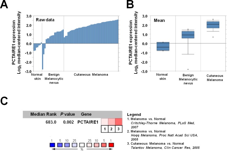
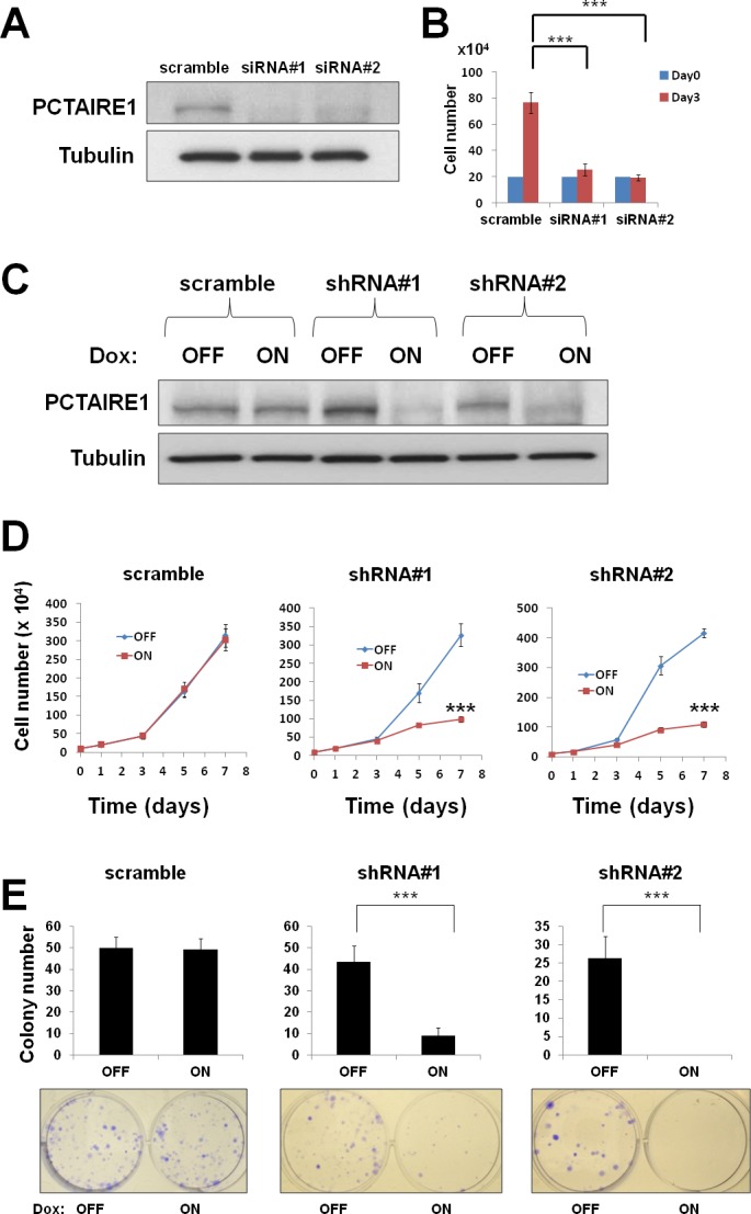
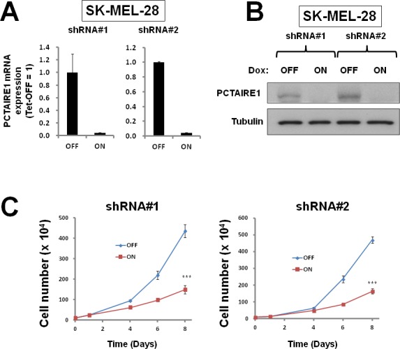
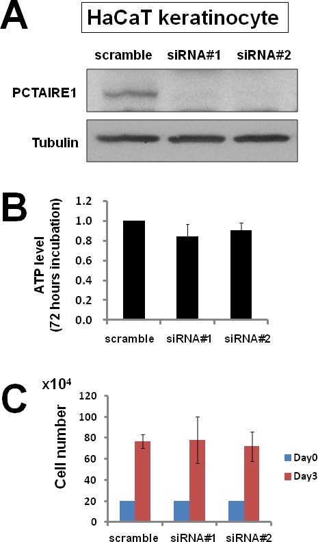
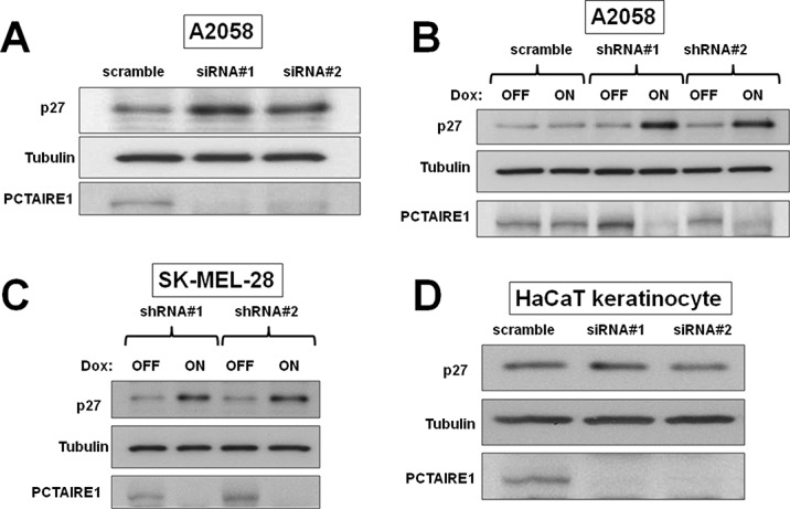
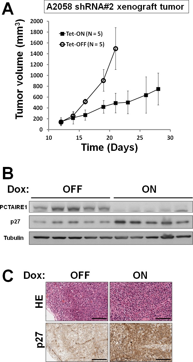
PCTAIRE1 is a cyclin-dependent kinase family protein that has been implicated in spermatogenesis. Although we recently revealed the function of PCTAIRE1 in tumorigenesis of epithelial carcinoma cells, its tumorigenic function in melanoma remains unclear. Interrogation of the Oncomine database revealed that malignant melanoma showed up-regulation of PCTAIRE1 mRNA compared to normal skin and benign melanocytic nevus tissues. In the melanoma cell lines A2058 and SK-MEL-28, PCTAIRE1 gene knockdown using siRNA or shRNA diminished melanoma cell proliferation as assessed by cellular ATP levels, cell counting and clonogenic assays. Moreover, FACS analyses of annexin V-PI staining and DNA content showed that PCTAIRE1 knockdown caused apoptosis in A2058 cells. In contrast, PCTAIRE1 does not appear to be involved in the proliferation of immortalized human keratinocyte HaCaT cells. Depletion of PCTAIRE1 by siRNA/shRNA led to p27 accumulation in melanoma cells but not HaCaT cells. In tumor xenografts of melanoma A2058 cells, conditional knockdown of PCTAIRE1 restored p27 protein expression and suppressed tumor growth. Our findings reveal a crucial role for PCTAIRE1 in regulating p27 protein levels and tumor growth in melanoma cells, suggesting that PCTAIRE1 could provide a target for melanoma treatment.

Keywords: PCTAIRE1; apoptosis; melanoma; p27.


Figures

Figure 1
Figure 1. PCTAIRE1 expression is up-regulated in melanomas
(A, B) Microarray datasets were accessed using the Oncomine database (http://www.oncomine.com). PCTAIRE1 expression (as log2 median-centered intensity) for normal skin, benign melanocytic nevus and malignant melanoma is shown either as raw data (A) or as the mean (B). In the chart showing the mean, dots indicate extreme data values. Melanoma vs. Normal: Fold change = 5.027, P = 3.98 × 10⁻⁹, Melanoma vs. Melanocytic nevus: Fold change = 2.504, P = 4.55 × 10⁻⁵. (C) Meta-analysis of PCTAIRE1 gene expression profiling where colored squares indicate the median rank for PCTAIRE1 (vs. Normal skin) across each analysis. PCTAIRE1 ranks in the top 10% in two of three analyses. The P value is given for the median-rank analysis.
Figure 2
Figure 2. PCTAIRE1 knockdown diminished melanoma A2058 cell growth
(A) A2058 cells were transfected with scrambled RNA or one of two different siRNAs targeting PCTAIRE1 mRNA (siRNA#1, #2). After 72 hours, cell lysates were analyzed by immunoblotting. (B) To measure cell growth, 2.0 × 10⁵ cells transfected with the indicated siRNAs were seeded onto 60 mm diameter plates. After 72 hours, the numbers of cells were counted. *** p < 0.001 (C) Protein lysates were generated from A2058 cells stably containing inducible shRNAs targeting PCTAIRE1 (shRNA#1, #2) and scramble control cultured for 72 hours with or without 100 ng/ml doxycycline, and analyzed by immunoblotting. (D) A2058 cells (10.0 × 10⁴) stably containing inducible shRNAs were cultured in 60 mm diameter plates for 24 hours, then stimulated with (ON) or without (OFF) doxycycline. The numbers of cells were counted (mean ± SD; n = 3). *** p < 0.001 (E) A2058 cells stably containing inducible shRNAs were seeded at 300 cells per well in 60 mm dishes. After 24 hours, doxycycline was added. Colonies consisting of > 50 cells were enumerated on day 10. All data represent mean ± SD (n = 3). *** p < 0.001
Figure 3
Figure 3. PCTAIRE1 knockdown diminished SK-MEL-28 cell growth
(A) SK-MEL-28 cells stably containing inducible shRNAs targeting different sites on PCTAIRE1 mRNA (shRNA#1, #2) were cultured for 3 days with 100 ng/ml doxycycline (Dox). PCTAIRE1 mRNA levels were measured by qRT-PCR, with normalization relative to GADPH (mean ± SD; n = 2). (B) SK-MEL-28 cells with inducible shRNAs (#1, #2) were cultured for 3 days with (ON) or without (OFF) doxycycline. Protein lysates were generated and analyzed by immunoblotting. (C) SK-MEL-28 cells (5.0 × 10⁴) stably containing shRNAs were cultured for 24 hours, then stimulated with (ON) or without (OFF) doxycycline. The numbers of cells were counted (mean ± SD; n = 3). *** p < 0.001
Figure 4
Figure 4. PCTAIRE1 knockdown did not diminish HaCaT keratinocyte growth
(A) HaCaT keratinocytes were transfected with scrambled RNA or two different siRNAs targeting PCTAIRE1 (siRNA#1, #2). Three days after transfection, cell lysates were prepared and analyzed by immunoblotting. (B) HaCaT cells were transfected with siRNAs as indicated. After 3 days, cellular ATP levels were measured using Cell Titer Glo reagents (mean ± SD; n = 3). (C) To measure cell growth, 2.0 × 10⁵ cells transfected with the indicated siRNAs were plated. After 3 days, the number of cells was counted (mean ± SD; n = 3).
Figure 5
Figure 5. PCTAIRE1 knockdown resulted in apoptosis of A2058 cells
(A, B) A2058 cells were transfected with the indicated siRNAs. Cells stained with annexin V and PI were assessed by FACS analyses at the indicated times. (A) Representative data at 72 hours after transfection are shown. (B) The percentage of apoptotic (annexin V positive and PI negative) cells is shown. All data represent mean ± SD (n = 3). *** p < 0.001 (C) A2058 cells transfected with siRNAs were cultured for 72 hours, followed by FACS analysis. Data represent relative DNA (propidium iodide fluorescence; x-axis) versus relative cell number (y-axis).
Figure 6
Figure 6. PCTAIRE1 knockdown leads to accumulation of p27
(A) A2058 cells were transfected with control RNA or two different siRNAs targeting PCTAIRE1. Three days after transfection, cell lysates were prepared and analyzed by immunoblotting. (B) A2058 cells stably containing inducible shRNAs targeting PCTAIRE1 (shRNA#1, #2) were cultured for 3 days with (ON) or without (OFF) 100 ng/ml doxycycline (Dox). Protein lysates were analyzed by SDS-PAGE/immunoblotting. (C) SK-MEL-28 cells with inducible shRNAs (#1, #2) were cultured for 3 days with (ON) or without (OFF) doxycycline (Dox). Protein lysates were analyzed by SDS-PAGE/ immunoblotting. (D) HaCaT keratinocytes were transfected with RNAs as indicated. After 3 days, cell lysates were prepared and analyzed by immunoblotting.
Figure 7
Figure 7. In vivo tumor xenograft analysis
(A) A2058 cells (5.0 × 10⁶) stably containing shRNA#2 were subcutaneously injected into the flanks of nu/nu mice. When tumor volumes reached 150 ~ 200 mm3 (Day 12), the animals were provided water with (white circles) or without (black square) 2% doxycycline (“Dox”) (n = 5 mice per group). Tumor volumes (mm3) were measured every other day (mean ± SD; n = 5). (B) Animals were sacrificed and tumors extracted for immunoblot analyses. (C) Representative examples of hematoxylin-eosin and p27 immunostaining are provided. Scale Bar = 100 μm.
Figure 8
Figure 8. Model of PCTAIRE1 function in melanomas
PCTAIRE1 promotes degradation of p27, thereby decreasing apoptosis of melanoma cells.

References

    1. Liu D, Liu X, Xing M. Activities of multiple cancer-related pathways are associated with BRAF mutation and predict the resistance to BRAF/MEK inhibitors in melanoma cells. Cell Cycle. 2014;13(2):208–219. - PMC - PubMed
    1. Saranga-Perry V, Ambe C, Zager JS, Kudchadkar RR. Recent developments in the medical and surgical treatment of melanoma. CA Cancer J Clin. 2014;64(3):171–185. - PubMed
    1. Vultur A, Herlyn M. SnapShot: melanoma. Cancer Cell. 2013;23(5):706–706. e701. - PubMed
    1. Shtivelman E, Davies MQ, Hwu P, Yang J, Lotem M, Oren M, Flaherty KT, Fisher DE. Pathways and therapeutic targets in melanoma. Oncotarget. 2014;5(7):1701–1752. - PMC - PubMed
    1. Cole AR. PCTK proteins: the forgotten brain kinases? Neurosignals. 2009;17(4):288–297. - PubMed