. 2014 Aug 21;1(8):513-521.
doi: 10.18632/oncoscience.74. eCollection 2014.

Rac1 as a multifunctional therapeutic target to prevent and combat cancer metastasis

Affiliations

Rac1 as a multifunctional therapeutic target to prevent and combat cancer metastasis

Christoph R Arnold et al. Oncoscience. .

Abstract

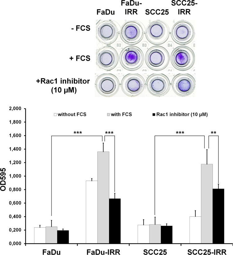
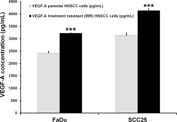
Metastatic progression of malignant tumors resistant to conventional therapeutic approaches is an ultimate challenge in clinical oncology. Despite the efforts of basic and clinical researchers, there is still no effective treatment schedule to prevent or combat metastatic spread of malignant tumors. This report presents recent findings that could help in the development of targeted therapeutics directed against the most aggressive and treatment-resistant carcinoma cells. It was demonstrated that HNSCC carcinoma cell lines with acquired treatment resistance possessed increased number of cells with carcinoma stem cell (CSC) properties. Furthermore, resistant cells were characterized by increased expression of Rac1, enhanced cell migration, and accelerated release of proangio- and vasculogenic factors (VEGF-A) and influence on endothelial cell (HMEC-1) migration. Inhibition of Rac1 signaling in the treatment-resistant carcinoma cells can interrupt metastatic process due to anoikis restoration and decrease of cell migration. It is also suggested that carcinoma cells with repressed survival capacities will be characterized by reduced release of proangiogenic factors, resulting in the decrease of endothelial cell migration. Therefore targeting of Rac1-related pathways may be considered as a promising therapeutic approach to prevent or combat metastatic lesions.

Keywords: Rac1; carcinoma stem cells; head and neck squamous cell carcinoma; metastasis; treatment resistance.


Conflict of interest statement

Conflict of Interest Statement

The authors indicate no potential conflicts of interest.

Figures

Figure 1
Figure 1. Notch-1 expression in HNSCC cell lines
Notch-1 expression in the parental treatment-sensitive and treatment-resistant IRR HNSCC cells was determined by ELISA (Notch-1 ELISA Kit, RayBiotech, Inc, Norcross, GA, USA) as described in the manufacturer's instructions. *p < 0.05
Figure 2
Figure 2. Protein patterns in radioresistant FaDu-IRR cells
(A) Proteins differently expressed in parental and treatment-resistant FaDu cells were identified by two-dimensional differential gel electrophoresis (2-D DIGE) followed by MALDI-TOF/TOF mass spectrometry. (B) Identified proteins were evaluated for their common targets and activated processes in radioresistant HNSCC cells. Visual representation of molecular networks of DIGE- and software-found proteins and cell processes was performed using two versions of the software PathwayStudio 8.0 (Ariadne Genomics Inc.) [9] and PathwayStudio 10.3 (Elsevier B.V., Amsterdam, The Netherlands) based on valuable Medline citations from various investigators. Key for the represented shapes: ellipse – identified proteins; rectangles – cell processes. Green ellipses indicate DIGE-identified down-regulated proteins, red ellipses represent up-regulated proteins, orange ellipses indicate software-found proteins. Grey arrows show the relationship between cell processes and identified proteins [9].
Figure 3
Figure 3. Modulation of expression of radiation-associated proteins in HNSCC cells
Exponentially growing HNSCC cells were collected for Western blot analysis. Total protein extracts were prepared from the cells and then processed for immunoblotting using antibodies to detect Rac1, PTEN (Cell Signaling Technology, Beverly, MA, USA), E-cadherin, and Akt/PKB (Santa Cruz Biotechnology, Inc., Santa Cruz, CA, USA). α-tubulin was used as a loading control.
Figure 4
Figure 4. Receptor status (EGFR and ErbB2, ErbB3) in radio-resistant HNSCC cells
Exponentially growing HNSCC carcinoma cells were analysed for expression of ErbB family members using RayBio® Human EGFR Phosphorylation Antibody Array 1 Kit (RayBiotech, Inc, Norcross, GA, USA). Integrated density values (IDVs) were normalized against the signal intensities of positive controls after background correction. Columns represent the mean value including standard deviation obtained from three independent experiments (*p < 0.05; **p < 0.01; ***p < 0.001).
Figure 5
Figure 5. Direct relationships between proteins of interest Rac1, Notch-1, c-myc, E-cadherin (CDH1), and PTEN
This network consisting of the analysed proteins was created using online software PathwayStudio 10.3 (Elsevier B.V., Amsterdam, The Netherlands) based on the Medline abstracts and full text publications.
Figure 6
Figure 6. Rac1 inhibitor enhances the sensitivity of HNSCC to ionizing radiation or cisplatin
(A) Parental FaDu, SCC25 and treatment-resistant IRR cells were exposed to ionizing radiation at a single dose of 2 Gy, to Rac1 inhibitor (20 μM) or to a combination of irradiation (2 Gy) and Rac1 inhibitor (20 μM). (B) parental and IRR HNSCC cells were treated with cisplatin at a clinically relevant single dose of 10 μM, with Rac1 inhibitor (20 μM) or with their combination, and then incubated for 72 hours. Cell viability and number of cells was evaluated using a Beckman Coulter Vi-CELL AS cell viability analyzer. Data are given as mean and standard deviation obtained from at least three independent experiments. *p<0.05, **p<0.01 - significance of differences in cell viability in HNSCC cells treated with ionizing radiation or cisplatin compared to their combinations wit Rac1 inhibitor [3].
Figure 7
Figure 7. Effects of Rac1 inhibitor on HNSCC cell migration
Differences in migration of parental and IRR HNSCC cells, and Rac1 inhibitor-induced repression of cell migration, were determined using a QCMTM 24-well colorimetric cell migration assay (Merck Millipore, Darmstadt, Germany), following the manufacturer's instructions. HNSCC cells harvested in the appropriate serum-free quenching medium were placed in the upper insert with an 8-μm pore size polycarbonate membrane. The lower chamber contained culture medium with chemoattractant (10% FCS). Plates were incubated for 24 hours at 37°C in a 5% CO2 humidified atmosphere. HNSCC cells that migrated through the membrane were stained and then subsequently extracted using extraction buffer. The optical densities of dye extracts were read at 560 nm using a microplate reader (Bio-Rad Microplate Reader 680, Bio-Rad Laboratories GmbH, Munich, Germany). **p < 0.01; ***p < 0.001 [9].
Figure 8
Figure 8. Assessment of VEGF-A concentrations in HNSCC cells
VEGF-A concentrations were determined by ELISA (RayBiotech, Inc, Norcross, GA, USA) in the secretomes obtained from parental and treatment-resistant HNSCC cells. Briefly, HNSCC cells were seeded into the T75 flasks and incubated overnight. Then cells were washed and incubated in serum-free culture medium. Twenty-four hours later, serum-starved medium was collected for the evaluation of VEGF-A levels as described in the manufacturer's instructions. Results are shown as mean and standard deviation obtained from three independent experiments. ***p < 0.001.
Figure 9
Figure 9. HMEC-1 cell migration toward secretomes collected from treatment-sensitive and treatment-resistant HNSCC cells
Secretomes from parental and IRR HNSCC cells were collected as described in the legend for Figure 7. Migration of HMEC-1 cells was assessed using a QCMTM 24-well colorimetric cell migration assay (Merck Millipore, Darmstadt, Germany) as described in Figure 7. The lower chamber contained HNSCC secretomes as a chemoattractant. The optical densities of dye extracts obtained from the migrated HMEC-1 cells were read at 560 nm using a microplate reader. **p < 0.01; ***p < 0.001.

References

    1. Haddad RI, Shin DM. Recent advances in head and neck cancer. N Engl J Med. 2008;359:1143–1154. - PubMed
    1. Begg AC. Predicting recurrence after radiotherapy in head and neck cancer. Semin. Radiat Oncol. 2012;22:108–118. - PubMed
    1. Skvortsov S, Dudas J, Eichberger P, Witsch-Baumgartner M, Loeffler-Ragg J, Pritz C, Schartinger VH, Maier H, Hall J, Debbage P, Riechelmann H, Lukas P, Skvortsova I. Rac1 as a potential therapeutic target for chemo-radioresistant head and neck squamous cell carcinomas (HNSCC) Br J Cancer. 2014;110:2677–2687. - PMC - PubMed
    1. Argiris A, Li Y, Forastiere A. Prognostic factors and long-term survivorship in patients with recurrent or metastatic carcinoma of the head and neck. Cancer. 2004;101:2222–2229. - PubMed
    1. Ferrari D, Codeca C, Fiore J, Luciani A, Foa P. A review on the treatment of relapsed/metastatic head and neck cancer. Expert Opin Pharmacother. 2009;10:2625–2632. - PubMed