. 2015 Mar;28(3):353-66.
doi: 10.1002/nbm.3258. Epub 2015 Jan 16.

A model-based reconstruction for undersampled radial spin-echo DTI with variational penalties on the diffusion tensor

Affiliations

A model-based reconstruction for undersampled radial spin-echo DTI with variational penalties on the diffusion tensor

Florian Knoll et al. NMR Biomed. 2015 Mar.

Abstract

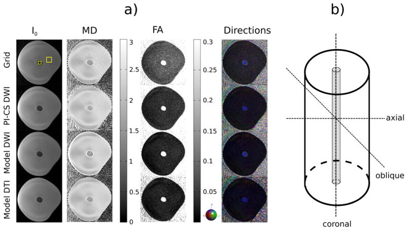
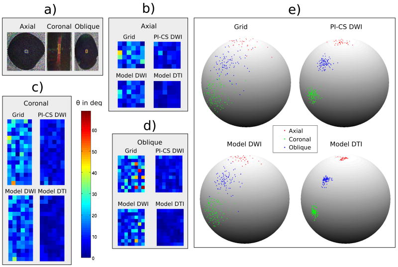
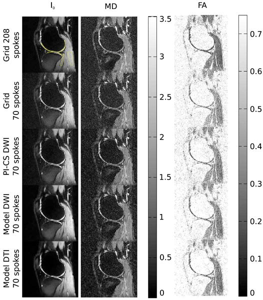
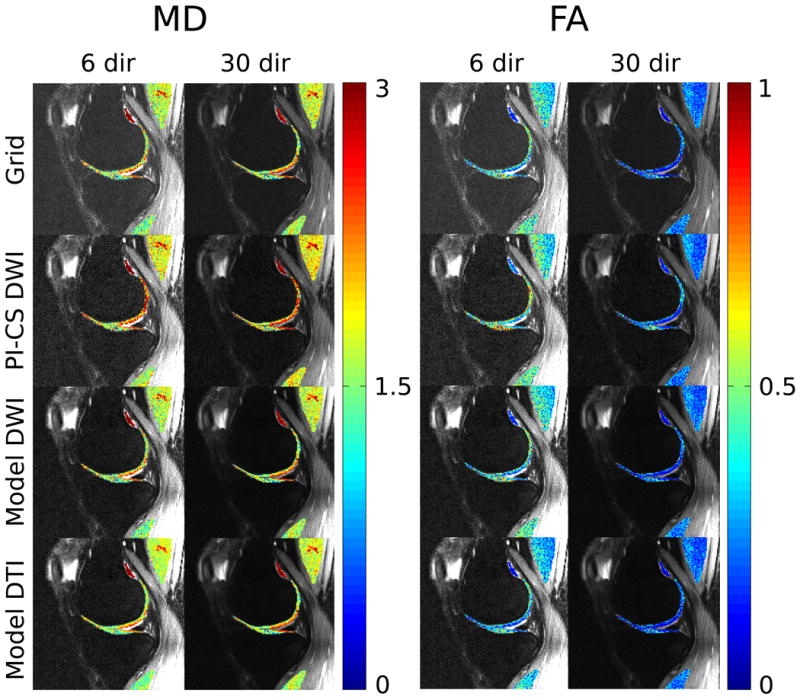
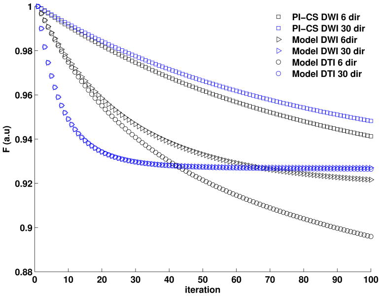
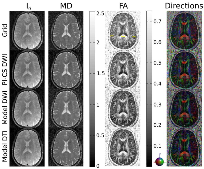
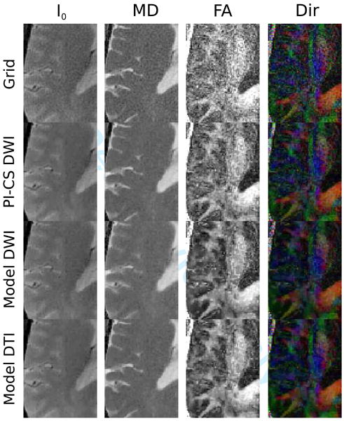
Radial spin-echo diffusion imaging allows motion-robust imaging of tissues with very low T2 values like articular cartilage with high spatial resolution and signal-to-noise ratio (SNR). However, in vivo measurements are challenging, due to the significantly slower data acquisition speed of spin-echo sequences and the less efficient k-space coverage of radial sampling, which raises the demand for accelerated protocols by means of undersampling. This work introduces a new reconstruction approach for undersampled diffusion-tensor imaging (DTI). A model-based reconstruction implicitly exploits redundancies in the diffusion-weighted images by reducing the number of unknowns in the optimization problem and compressed sensing is performed directly in the target quantitative domain by imposing a total variation (TV) constraint on the elements of the diffusion tensor. Experiments were performed for an anisotropic phantom and the knee and brain of healthy volunteers (three and two volunteers, respectively). Evaluation of the new approach was conducted by comparing the results with reconstructions performed with gridding, combined parallel imaging and compressed sensing and a recently proposed model-based approach. The experiments demonstrated improvements in terms of reduction of noise and streaking artifacts in the quantitative parameter maps, as well as a reduction of angular dispersion of the primary eigenvector when using the proposed method, without introducing systematic errors into the maps. This may enable an essential reduction of the acquisition time in radial spin-echo diffusion-tensor imaging without degrading parameter quantification and/or SNR.

Keywords: compressed sensing; diffusion-tensor imaging; iterative reconstruction; model-based image reconstruction; non-Cartesian imaging.


Figures

Figure 1
Figure 1
a) Axial I0 image and maps of MD, FA and the color coded direction of the principal eigenvector of the phantom measurement with 61 spokes, two b-values and 6 diffusion-encoding directions for conventional gridding (Grid, first row), combined PI and CS on the diffusion-weighted images (PI-CS DWI, second row), model-based reconstruction with TV regularization of the diffusion-weighted images (Model DWI, third row) and model-based reconstruction with TV regularization of the tensor elements (Model DTI, fourth row). The ROIs used for the analysis in Table 1 are highlighted in the gridding reconstruction of the I0 image. b) Schematic of the phantom. The orientations where data was acquired are shown as dashed lines. The Dyneema fibers are located at the center of the phantom and are responsible for the anisotropy in the central region of the images in a.
Figure 2
Figure 2
Dispersion of the principal eigenvector in the anisotropic region of the phantom for three different orientations (axial, coronal and oblique at a 45 degree angle with respect to the anisotropic fibers). While all nonlinear methods show a clear reduction of dispersion the effect is strongest for the proposed Model DTI approach. This behavior is consistent over the different orientations of the anisotropy. a) Color coded direction of the principal eigenvector for the three different orientations. b,c,d) Color coded deviations of the dispersion angle θ to the mean of all vectors in the ROI in degrees. e) Illustration of the dispersion of the eigenvectors in the ROIs.
Figure 3
Figure 3
I0 image and maps of MD and FA from measurements of the knee of a healthy volunteer with 208 and 70 spokes and two b-values for conventional gridding (Grid, first row 208 spokes, second row 70 spokes), combined PI and CS on the diffusion-weighted images (PI-CS DWI, third row), model-based reconstruction with TV of the diffusion-weighted images (Model DWI, fourth row) and model-based reconstruction with TV of the tensor elements (Model DTI, fifth row). The ROIs that were used for the evaluation of cartilage and muscle tissue in Table 2 are highlighted in the 208 spokes gridding reconstruction of the I0 image.
Figure 4
Figure 4
I0 images with overlaid maps of MD and FA in cartilage and muscle from measurements of the knee of a healthy volunteer with 70 spokes using 6 and 30 directions for conventional gridding (Grid, first row), combined PI and CS on the diffusion-weighted images (PI-CS DWI, second row), model-based reconstruction with TV of the diffusion-weighted images (Model DWI, third row) and model-based reconstruction with TV of the tensor elements (Model DTI, fourth row). Quantitative values are presented in Table 3.
Figure 5
Figure 5
Convergence plots of the different iterative reconstruction methods from Fig. 4. The convergence of Model-DWI and Model-DTI are very similar, and indicate proper convergence of the methods while convergence of PI-CS is slower.
Figure 6
Figure 6
I0 image and maps of MD, FA and the color coded direction of the principal eigenvector from 196-matrix 75 spokes brain measurements for conventional gridding (Grid, first row), combined PI and CS on the diffusion-weighted images (PI-CS DWI, second row), model-based reconstruction with TV of the diffusion-weighted images (Model DWI, third row) and model-based reconstruction with TV of the tensor elements (Model DTI, fourth row). The ROIs used for the analysis of the splenium of the corpus callosum and grey matter in Table 4 are highlighted in the gridding reconstruction of the FA map.
Figure 7
Figure 7
An enlarged region from brain acquisitions in Figure 6. Note the pronounced reduction of noise in FA and direction color maps for all nonlinear reconstructions, which is strongest for the two model based methods. In the case of Model DWI this comes at the cost of blurring and a loss of fine details in the MD maps, an effect that does not occur in Model DTI.

References

    1. Tournier JD, Mori S, Leemans A. Diffusion tensor imaging and beyond. Magn Reson Med. 2011;65(6):1532–1556. - PMC - PubMed
    1. Maroudas A, Bayliss MT, Uchitel-Kaushansky N, Schneiderman R, Gilav E. Aggrecan turnover in human articular cartilage: use of aspartic acid racemization as a marker of molecular age. Arch Biochem Biophys. 1998;350(1):61–71. - PubMed
    1. Verzijl N, DeGroot J, Thorpe SR, Bank RA, Shaw JN, Lyons TJ, Bijlsma JW, Lafeber FP, Baynes JW, TeKoppele JM. Effect of collagen turnover on the accumulation of advanced glycation end products. J Biol Chem. 2000;275(50):39027–39031. - PubMed
    1. Lesperance LM, Gray ML, Burstein D. Determination of fixed charge density in cartilage using nuclear magnetic resonance. J Orthop Res. 1992;10(1):1–13. - PubMed
    1. Bashir A, Gray ML, Burstein D. Gd-DTPA2- as a measure of cartilage degradation. Magn Reson Med. 1996;36(5):665–673. - PubMed

Publication types

LinkOut - more resources