. 2015 Jan 15;160(1-2):177-90.
doi: 10.1016/j.cell.2014.12.019.

Glial lipid droplets and ROS induced by mitochondrial defects promote neurodegeneration

Affiliations

Glial lipid droplets and ROS induced by mitochondrial defects promote neurodegeneration

Lucy Liu et al. Cell. .

Abstract

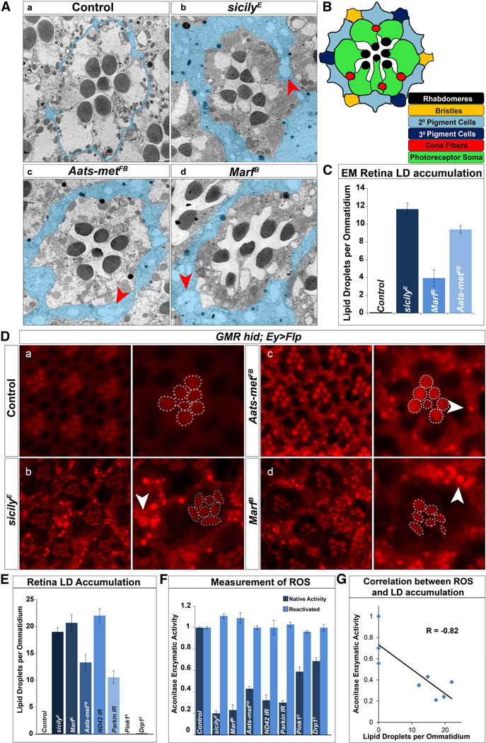
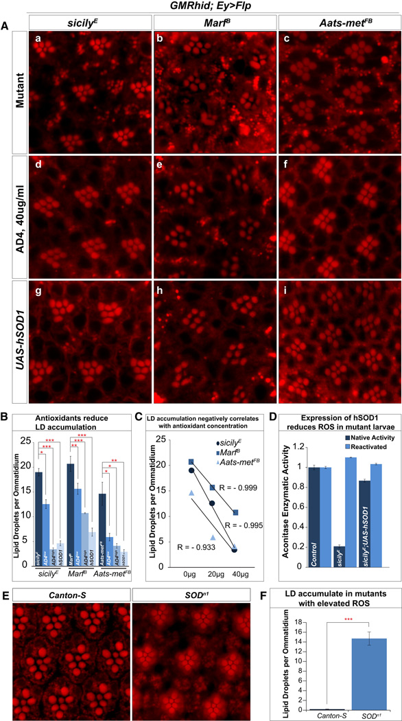
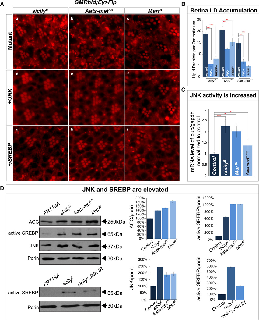
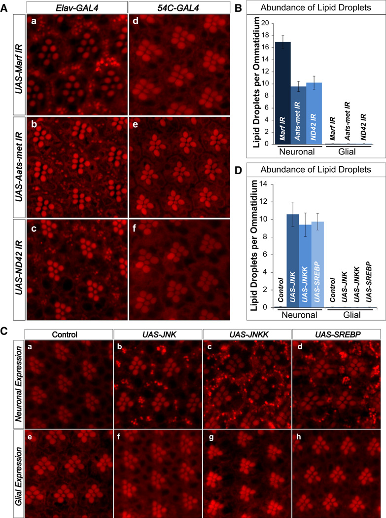
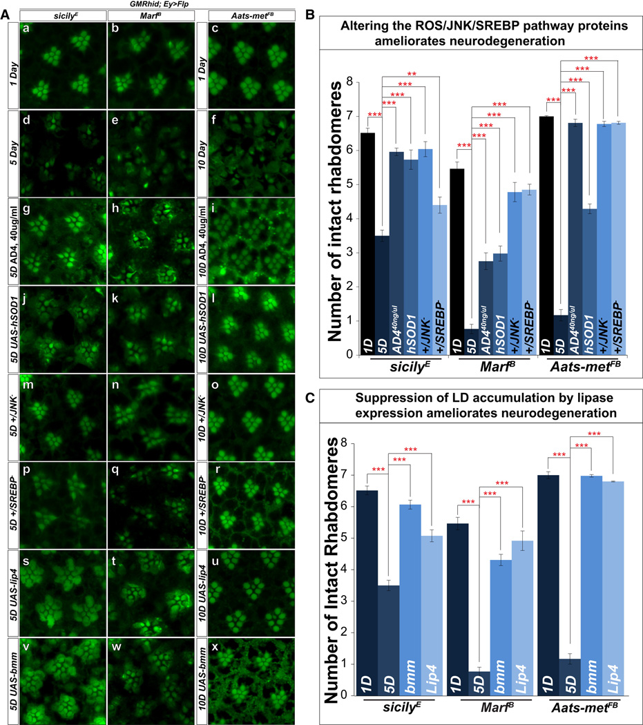
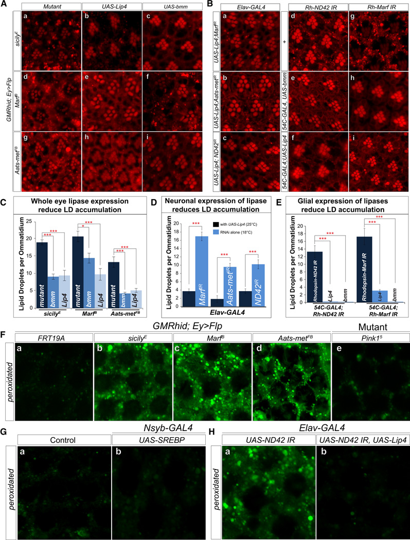
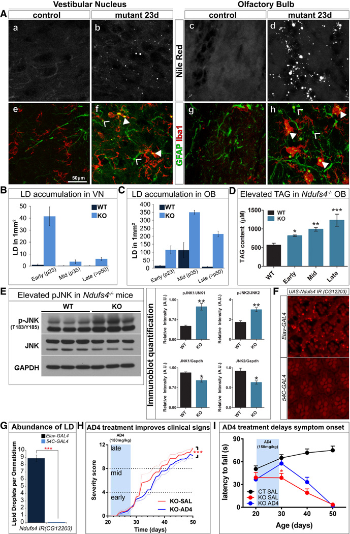
Reactive oxygen species (ROS) and mitochondrial defects in neurons are implicated in neurodegenerative disease. Here, we find that a key consequence of ROS and neuronal mitochondrial dysfunction is the accumulation of lipid droplets (LD) in glia. In Drosophila, ROS triggers c-Jun-N-terminal Kinase (JNK) and Sterol Regulatory Element Binding Protein (SREBP) activity in neurons leading to LD accumulation in glia prior to or at the onset of neurodegeneration. The accumulated lipids are peroxidated in the presence of ROS. Reducing LD accumulation in glia and lipid peroxidation via targeted lipase overexpression and/or lowering ROS significantly delays the onset of neurodegeneration. Furthermore, a similar pathway leads to glial LD accumulation in Ndufs4 mutant mice with neuronal mitochondrial defects, suggesting that LD accumulation following mitochondrial dysfunction is an evolutionarily conserved phenomenon, and represents an early, transient indicator and promoter of neurodegenerative disease.


Figures

Figure 1
Figure 1. LD accumulate in glia of mitochondrial mutants with elevated levels of reactive oxygen species (ROS)
TEM of a single ommatidium of 1-day-old flies. A) a, Pigment cells that surround the photoreceptors are typically very thin (blue) in wild type eyes (y w FRT19A clones). They are vastly expanded in b, sicily, c, Aats-met, and d, Marf mutants and contain numerous LD (red arrowhead). B) Schematic of a single ommatidium in cross-section. C) Quantification of retina LD per ommatidium. D) a–d, Nile Red stained whole-mount retina show LD accumulation (white arrowhead) in sicily, Marf and Aats-met mutant clones of 1-day-old flies, but not in controls (y w FRT19A clones). Rhabdomeres outlined in white. Abnormal rhabdomere morphology can be seen in sicily and Marf mutant clones. E) Quantification of LD per ommatidium of panel D. A minimum of 16 eyes were stained and approximately 36 ommatidia were quantified. F) Aconitase activity is used to measure ROS - before and after enzymatic reactivation with reducing agents. RNAis were driven by the ubiquitously expressed daughterless[da]-GAL4 G) Correlation between aconitase enzymatic activity and LD per ommatidium. R = correlation coefficient. Data are represented as mean ± SEM. See also figure S1–3.
Figure 2
Figure 2. Antioxidants reduce LD accumulation in mutant retinas
A) a–c, Nile Red stains of 1-day-old sicily, Marf and Aats-met mutant retinas reveal LD accumulation. d–f, Animals fed with AD4 at 40µg/ml (AD4high in quantification) reduces the number of LD. Notice the partial restoration of rhabdomere integrity in Marf mutants. g–i, Suppressing ROS by overexpression of a copy of hSOD1 (eyeless[ey]-Gal4, UAS-Flp) also reduces LD accumulations. B) Quantification of the data presented in A and animals with mutant clones raised on food supplemented with 20µg/ml (AD4low) of AD4 the show a reduction of LD accumulation. C) Linear regressions show a negative correlation between the numbers of LD accumulated and the amount of supplemented AD4. D) Expression of hSOD1 with da-Gal4 in sicily mutants significantly suppresses the loss of aconitase activity and hence ROS E) Nile Red staining of wild type (Canton-S) flies show no LD accumulation whereas staining of SODn1 mutant escapers reveal high levels of LD accumulation. F) Quantification of SODn1 LD accumulation. Data are represented as mean ± SEM. Significance was calculated compared to controls using t-tests (*P<0.05, **P<0.005, ***P<0.0005).
Figure 3
Figure 3. ROS leads to aberrant activation of JNK and SREBP and reducing JNK or SREBP in mutants reduces LD accumulation
A) a–c, sicily, Aats-met and Marf mutant clones exhibit high levels of LD accumulation but when one copy of JNK (d–f) or SREBP (g–i) is removed, LD accumulation is significantly reduced. B) Quantification of retinas in A. C) qRT-PCR quantification of puc shows a significant upregulation of puc mRNA in all three mutant larvae compared to control (y w FRT19A). D) Immunoblot of sicily, Aats-met and Marf heads with mutant eye clones show an increase in ACC, total JNK and active SREBP when compared to control (y w FRT19A). Knockdown of JNK in the sicily mutant background (heads), reduces active SREBP levels compared to heads with mutant visual systems. Data are represented as mean ± SEM. Significance was calculated compared to controls and immunoblots were normalized to loading control (porin). (*P<0.05, **P<0.005, ***P<0.0005). See also Figure S4.
Figure 4
Figure 4. Photoreceptor neurons, but not glia, induce LD accumulation cell non-autonomously
A) a–c, Nile Red stains show that neuronal knockdown (elav-GAL4) of Marf, Aats-met, and ND42 result in LD accumulation in the glia. d–f, Glial (54C-GAL4) knockdown of Marf, Aats-met, and ND42 does not result in LD accumulation. B) Quantification of A. C) a,e, Control (white) do not exhibit LD accumulation. b–d, Overexpression of JNK, JNKK and SREBP in the neurons (elav-GAL4) leads to LD accumulation in the glia. e–h, However, overexpression of JNK, JNKK and SREBP in the glia (54C-GAL4) does not result in LD accumulation. D) Quantification of LD abundance. Data are represented as mean ± SEM. See also Figure S5.
Figure 5
Figure 5. Reduction of LD accumulation restores rhabdomere integrity and partially suppresses neurodegeneration
A) a–c, Phalloidin staining of whole mount retina of 1-day-old sicily, Marf and Aats-met mutant clones exhibit mostly intact rhabdomeres at this early stage. d–f, Upon aging, sicily (5d), Marf (5d), and Aatsmet (10d) mutant clones show obvious signs of degeneration. g–I, animals fed with 40µg/ml of AD4 exhibit more intact rhabdomeres after aging for all three mutants. j–l, Reducing LD accumulation by overexpression of hSOD1, or m–r, one copy of JNK or SREBP all partially restore rhabdomere integrity after aging based on phalloidin staining. s–x, The expression of lipases (brummer and Lip4) in the mutant background also delays degeneration compared to mutant clones alone. B) Quantification of rhabdomere numbers after aging show that expression of hSOD1, feeding AD4 and removing one copy of JNK or SREBP restores rhabdomere integrity. C) Quantification of rhabdomere numbers after aging shows that expression of both brummer and Lip4 significantly restore the number of photoreceptors in all three mutant backgrounds. Data are represented as mean ± SEM. Significance was calculated compared to controls (*P<0.05, **P<0.005, ***P<0.0005). See also Figure S6.
Figure 6
Figure 6. Lipid peroxidation in glial cells affects the demise of neurons
A)a–i, Nile Red staining of whole mount retina show that overexpression of brummer and Lip4 in the visual system reduces LD accumulations in sicily, Marf and Aats-met mutant clones. B) a–c, Neuronal expression of lipases and knockdown of Marf, Aats-met and sicily also reduces LD accumulation in the glia. Flies with elav-GAL4 mediated knockdown of the RNAis alone were raised at 18°C (efficient RNAi leads to high percentage of death) and flies expressing Lip4 and RNAi were raised at 25°C. d–i, Rh-ND42 and Marf RNAi in the neurons results in glial LD accumulation. Expression of brummer and Lip4 in the glia (54C-GAL4) reduces the formation of LD accumulation. C) Quantification of A. D) Quantification of B a–c, Significance was calculated compared to Figure 4Aa–c. E) Quantification of B d–i. F) a, e. Measurement of peroxidated lipids shows basal level of peroxidated lipids in control (FRT19A) clones and pink15 mutant retinas. b–d, 1-day-old mutant clones exhibit elevated levels of peroxidated lipids. G) Neuronal overexpression of SREBP does not result in elevated levels of peroxidated lipids compared to control (UAS-SREBP). H) Neuronal RNAi knockdown of ND42 results in elevated levels of peroxidated lipids, which is rescued with neuronal expression of lipase. Data are represented as mean ± SEM. Significance was calculated compared to controls. (*P<0.05, **P<0.005, ***P<0.0005). See also Figure S6.
Figure 7
Figure 7. LD accumulation is present in the astrocytes and microglia of Ndufs4−/− mice prior to neurodegeneration
A) a–d 20µm coronal cryosections reveal LD accumulation in the VN and OB of Ndufs4−/− mice compared to control (C57Bl/6). e–h LD accumulates in the astrocytes and microglia of the VN and OB shown by colocalization with the astrocyte marker, GFAP and the microglia marker, Iba1. (B) LD accumulation in the VN is high in the P23 mutants but decreases in number in the P34 and >P50 mutants. (C) In the OB, LD accumulation occurs in the P23 mutants and increases in number in the P34 and >P50 animals. (D) Ndufs4−/− exhibit elevated triglyceride (TAG) levels in the OB in an age-dependent manner. E) pJNK is increased in the mutants. Right panels show the quantification of the immunoblots. F) Neuronal knockdown of dNdufs4 (CG12203) leads to LD accumulation in the glia, while glial knockdown results in no LD accumulation. (*P<0.05, **P<0.005, ***P<0.0005). G) Quantification of F. H) 7-day administration of AD4 in P21 animals via IP injection at 150mg/kg shows treated mutant animals with improved clinical signs (body weight loss, hypotonia, ataxia, piloerection, clasping, gasping, paralysis, tremor) upon aging and I) performing at a comparable level as control while mutants treated with saline show progressive decline in motor performance. (*p<0.05 KO SAL vs KO AD4, two-way ANOVA). See also Figure S7.

References

    1. Amer J, Atlas D, Fibach E. N-acetylcysteine amide (AD4) attenuates oxidative stress in betathalassemia blood cells. Biochim. Biophys. Acta. 2008;1780:249–255. - PubMed
    1. Assouline Z, Jambou M, Rio M, Bole-Feysot C, de LP, Barnerias C, Desguerre I, Bonnemains C, Guillermet C, Steffann J, Munnich A, Bonnefont JP, Rotig A, Lebre AS. A constant and similar assembly defect of mitochondrial respiratory chain complex I allows rapid identification of NDUFS4 mutations in patients with Leigh syndrome. Biochim. Biophys. Acta. 2012;1822:1062–1069. - PubMed
    1. Bao S, Fischbach KF, Corbin V, Cagan RL. Preferential adhesion maintains separation of ommatidia in the Drosophila eye. Dev. Biol. 2010;344:948–956. - PMC - PubMed
    1. Barnham KJ, Masters CL, Bush AI. Neurodegenerative diseases and oxidative stress. Nat. Rev. Drug Discov. 2004;3:205–214. - PubMed
    1. Bayat V, Thiffault I, Jaiswal M, Tetreault M, Donti T, Sasarman F, Bernard G, Demers-Lamarche J, Dicaire MJ, Mathieu J, Vanasse M, Bouchard JP, Rioux MF, Lourenco CM, Li Z, Haueter C, Shoubridge EA, Graham BH, Brais B, Bellen HJ. Mutations in the mitochondrial methionyl-tRNA synthetase cause a neurodegenerative phenotype in flies and a recessive ataxia (ARSAL) in humans. PLoS. Biol. 2012;10:e1001288. - PMC - PubMed

Publication types

MeSH terms

Substances