. 2015 Jan 15;160(1-2):219-27.
doi: 10.1016/j.cell.2014.11.049.

Conformational changes of elongation factor G on the ribosome during tRNA translocation

Affiliations

Conformational changes of elongation factor G on the ribosome during tRNA translocation

Jinzhong Lin et al. Cell. .

Abstract

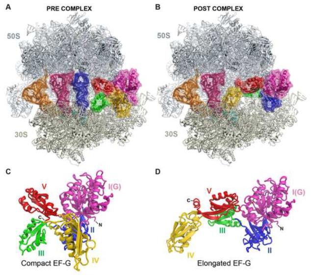
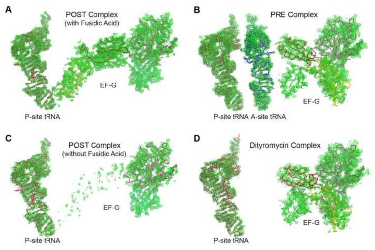
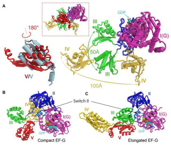
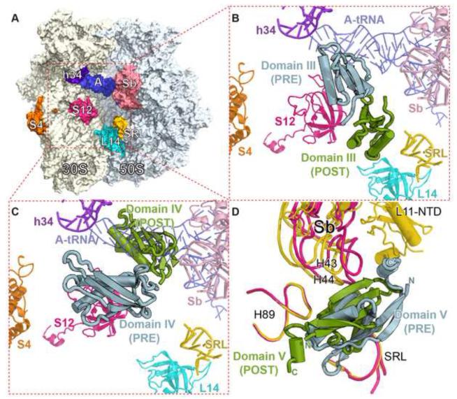
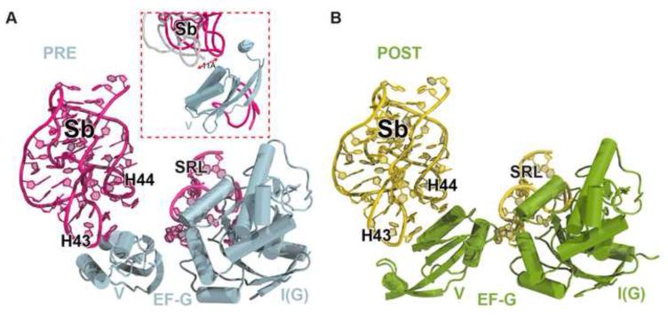
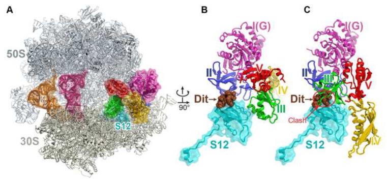
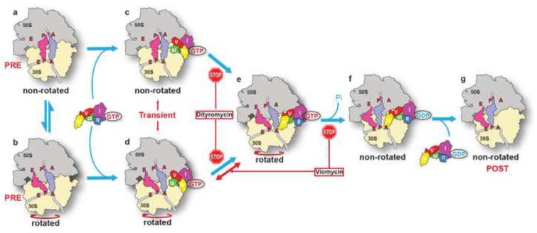
The universally conserved GTPase elongation factor G (EF-G) catalyzes the translocation of tRNA and mRNA on the ribosome after peptide bond formation. Despite numerous studies suggesting that EF-G undergoes extensive conformational rearrangements during translocation, high-resolution structures exist for essentially only one conformation of EF-G in complex with the ribosome. Here, we report four atomic-resolution crystal structures of EF-G bound to the ribosome programmed in the pre- and posttranslocational states and to the ribosome trapped by the antibiotic dityromycin. We observe a previously unseen conformation of EF-G in the pretranslocation complex, which is independently captured by dityromycin on the ribosome. Our structures provide insights into the conformational space that EF-G samples on the ribosome and reveal that tRNA translocation on the ribosome is facilitated by a structural transition of EF-G from a compact to an elongated conformation, which can be prevented by the antibiotic dityromycin.


Figures

Figure 1
Figure 1. The structures of EF-G bound to the pre- and post-translocation ribosome
(A and B) Overview of EF-G bound to the PRE (A) and the POST (B) ribosome. Shown are the 50S (gray) and the 30S (ivory) subunits, the A-site (blue), P-site (pink) and E-site (orange) tRNAs, mRNA (cyan) and EF-G with its five domains colored differently. (C and D) Cartoon representations of EF-G shown in the compact conformation (C) from the PRE complex and the elongated conformation (D) from the POST complex. Domains of EF-G are colored and labeled as in panels A and B. See also Figure S1, Table S1 and Movie S1.
Figure 2
Figure 2. Partial electron density for the POST and PRE complexes
(A-D) Unbiased FobsFcalc difference Fourier map of EF-G and the P-site tRNA in the POST complex in the presence of fusidic acid (A), EF-G and the A- and P-site tRNAs in the PRE complex (B), EF-G and the P-site tRNA in the POST complex in the absence of fusidic acid (C), and EF-G and the P-site tRNA in the dityromycin complex (D). All maps are contoured at 2.5σ obtained after initial refinement with an empty ribosome as a starting model. See also Table S1.
Figure 3
Figure 3. Comparison of the EF-G structures in the elongated and the compact conformations
(A) Inset is a superimposition of the structures of the compact and the elongated EF-G through domain I. Helices in the compact EF-G are displayed as cylinders. Lower right is a close-up view of the movements of domains III and IV. For clarity, conformational change of domain V is displayed separately in the lower left where the one from the compact EF-G is colored in lightblue. The GDP nucleotide is shown as spheres. (B and C) Structures of the compact (B) and the elongated (C) EF-G viewed from the catalytic site. The switch II is colored in lightblue as indicated. The switch I loop is disordered and not shown in both complexes. See also Movie S1 and Movie S2.
Figure 4
Figure 4. Interfaces between domains III, IV and V of EF-G and the ribosome in the PRE and the POST complexes
(A) Overview of the ribosome showing the orientation of the insets B and C. Elements of the ribosome are indicated and colored differently. (B and C) Positions of domains III (B) and IV (C) on the ribosome in the two complex structures. (D) Surroundings of domain V in the two complex structures. Helix 43/44, L11-NTD in the stalk base (Sb), Helix 89 and the sarcin-ricin loop (SRL) are shown in yellow in the POST complex and pink in the PRE complex. The L11-NTD in the PRE complex is not visible.
Figure 5
Figure 5. Contacts between domain V of EF-G, the stalk base (Sb) and the sarcin-ricin loop (SRL)
(A and B) Nucleotides at the tips of H43, H44 and the SRL are exposed in the PRE complex and are in close contacts with domain V in the POST complex. Positions of Sb from the two copies in the crystal of the PRE complex shown in the inset of panel A demonstrate that Sb becomes flexible in the PRE state. See also Figure S2.
Figure 6
Figure 6. The structure of EF-G bound to the ribosome trapped by the antibiotic dityromycin
(A) Overview of the structure of the complex. The components are colored in the same scheme as in Figure 1, except that here ribosomal protein S12, instead of mRNA, is shown in cyan. Dityromycin (Dit) binds to protein S12 located behind EF-G. (B) Close up of EF-G, S12 and dityromycin (brown). (C) Model of EF-G from the POST complex on the ribosome showing the steric collision between domain III and dityromycin. See also Movie S2.
Figure 7
Figure 7. Conformational changes of EF-G on the ribosome
EF-G in complex with GTP transiently folds into a compact conformation, from an elongated conformation in the ribosome-free state, after engaging the ribosome in the pre-translocational state to avoid a collision with the A-site tRNA (steps a-d). Rotation of the 30S subunit enables domain IV moving next to the A-site tRNA, a step that can be blocked by the antibiotic dityromycin (steps c-e). Further conformational changes of EF-G with concomitant GTP hydrolysis facilitate tRNA translocation (steps e-f). This process involves swiveling of the 30S head domain (Ramrath et al., 2013), which is not shown here. The antibiotic viomycin prevents translocation by locking the ribosome in the rotated state, while not affecting the initial conformational changes of EF-G (Munro et al., 2010). tRNA translocation is completed by dissociation of EF-G in complex with GDP from the ribosome (steps f-g).

References

    1. Adams PD, Grosse-Kunstleve RW, Hung LW, Ioerger TR, McCoy AJ, Moriarty NW, Read RJ, Sacchettini JC, Sauter NK, Terwilliger TC. PHENIX: building new software for automated crystallographic structure determination. Acta Crystallogr D Biol Crystallogr. 2002;58:1948–1954. - PubMed
    1. Agrawal RK, Heagle AB, Penczek P, Grassucci RA, Frank J. EF-G-dependent GTP hydrolysis induces translocation accompanied by large conformational changes in the 70S ribosome. Nat Struct Biol. 1999;6:643–647. - PubMed
    1. Agrawal RK, Penczek P, Grassucci RA, Frank J. Visualization of elongation factor G on the Escherichia coli 70S ribosome: the mechanism of translocation. Proc Natl Acad Sci U S A. 1998;95:6134–6138. - PMC - PubMed
    1. Blaha G, Stanley RE, Steitz TA. Formation of the first peptide bond: the structure of EF-P bound to the 70S ribosome. Science. 2009;325:966–970. - PMC - PubMed
    1. Blanchard SC, Kim HD, Gonzalez RL, Jr., Puglisi JD, Chu S. tRNA dynamics on the ribosome during translation. Proc Natl Acad Sci U S A. 2004;101:12893–12898. - PMC - PubMed

Publication types

MeSH terms