. 2015 Jan 15;160(1-2):241-52.
doi: 10.1016/j.cell.2014.12.032.

Hematopoietic stem cell arrival triggers dynamic remodeling of the perivascular niche

Affiliations

Hematopoietic stem cell arrival triggers dynamic remodeling of the perivascular niche

Owen J Tamplin et al. Cell. .

Abstract

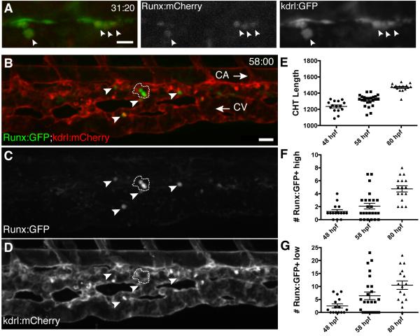
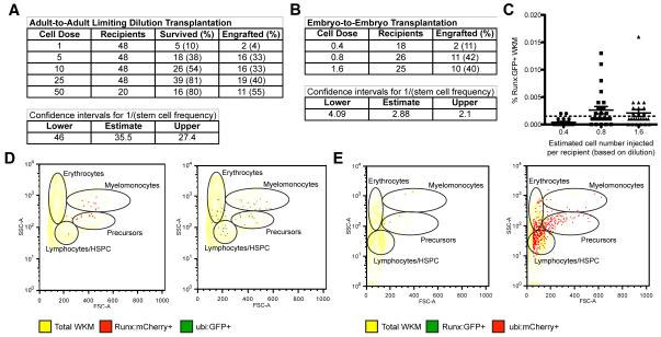
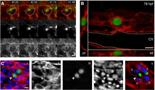
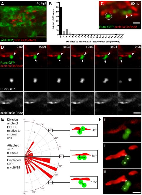
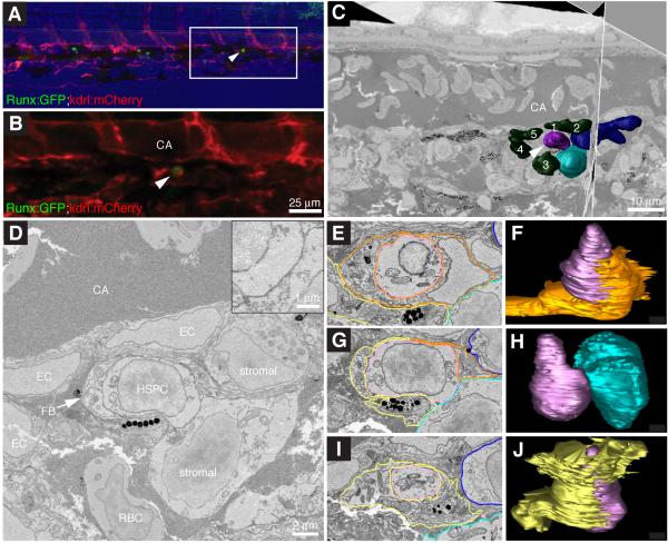
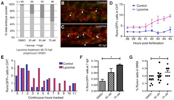
Hematopoietic stem and progenitor cells (HSPCs) can reconstitute and sustain the entire blood system. We generated a highly specific transgenic reporter of HSPCs in zebrafish. This allowed us to perform high-resolution live imaging on endogenous HSPCs not currently possible in mammalian bone marrow. Using this system, we have uncovered distinct interactions between single HSPCs and their niche. When an HSPC arrives in the perivascular niche, a group of endothelial cells remodel to form a surrounding pocket. This structure appears conserved in mouse fetal liver. Correlative light and electron microscopy revealed that endothelial cells surround a single HSPC attached to a single mesenchymal stromal cell. Live imaging showed that mesenchymal stromal cells anchor HSPCs and orient their divisions. A chemical genetic screen found that the compound lycorine promotes HSPC-niche interactions during development and ultimately expands the stem cell pool into adulthood. Our studies provide evidence for dynamic niche interactions upon stem cell colonization. PAPERFLICK:


Figures

Figure 1
Figure 1. A transgenic zebrafish line that specifically marks HSPCs
(A) Single frame of time-lapse (hours post fertilization:minutes) showing Runx:mCherry+ HSPCs (arrowheads; red nuclei) budding from kdrl:GFP+ hemogenic endothelium of DA (green). See Movie S1. (B-D) CHT is colonized by Runx:GFP+ HSPCs (green) that are closely associated with kdrl:mCherry+ ECs (red). Caudal artery (CA) is dorsal to CHT, caudal vein (CV) is ventral, circulation runs posterior (right arrow) and anterior (left arrow), respectively. Cluster of 3 Runx:GFP+ high cells outlined with dashed line and 4 Runx:GFP+ low cells indicated with arrowheads. 58 hpf embryo. (E-G) Runx:GFP+ high and low cells quantified, with CHT length as indicator of stage (anterior from cloaca to posterior limit of CA). Note: Confocal images are 3D rendered depth or max projections of 20-30 μm z-stacks. Scale bars: (A) 15 μm; (B) 25 μm. Error bars show mean ± SEM. See also Figure S1.
Figure 2
Figure 2. Functional HSPCs are highly purified using a single transgenic marker
Summary of results from (A) adult-to-adult and (B) embryo-to-embryo limiting dilution transplantation experiments. (C) Embryo-to-embryo transplantation recipients with engraftment of Runx:GFP+ cells in kidney marrow at 3 months (above background; >0.001%). Representative kidney marrow flow cytometry analysis of (D) adult-to-adult transplantation recipient with Runx:mCherry+ HSPCs and ubi:GFP+ lineages, and (E) embryo-to-embryo transplantation recipient with Runx:GFP+ HSPCs and ubi:mCherry+ lineages. Error bars show mean ± SEM. See also Figure S2.
Figure 3
Figure 3. Endothelial cells in the perivascular niche remodel to surround a single HSPC
(A) 4 frames from time-lapse Movie S2 between 40-42 hpf (hours post fertilization:minutes). Upper row is merge of Runx:GFP+ HSPC (green, middle row) and kdrl:RFP ECs (red, lower row). A group of surrounding ECs (broken circle) remodel around a single HSPC soon after its arrival. (B) Higher magnification (60x) live image of single Runx:GFP+ HSPC surrounded by kdrl:mCherry ECs at 78 hpf (orthogonal views). (C) 3D rendered projection of scanning confocal image from fixed 80 hpf embryo. (i) merge, (ii) kdrl:mCherry ECs, (iii) Runx:GFP+ HSPCs, (iv) DRAQ5 nuclei, (v) kdrl:mCherry projection with 3D modeled green HSPCs and 5 blue surrounding EC nuclei (arrowhead indicates HSPC in EC surround). All views: dorsal up, ventral down. Scale bars: 10 μm. See also Figure S3.
Figure 4
Figure 4. Endothelial cells surround HSPCs in the fetal liver microenvironment
(A-C) FLs from E11.5 Ly6a-GFP mice were fixed and stained for immunofluorescence with anti-VE-Cadherin (red), anti-Runx1 (blue), and anti-GFP (green) antibodies. We scored 59 Ly6a-GFP+/Runx1+ cells from 3 FLs and identified three different HSPC-EC configurations: (A) abluminal with no contact between HSPC and ECs (18/59; 30%); (B) EC contact on one side of the HSPC (27/59; 46%); (C) HSPC surrounded on all sides with ECs (14/59; 24%). See Movie S3. (D) c-kit+ cell (magenta) adhered to CD31+ ECs (green) in one lobe of an E11.5 FL (arrowhead). White box marks details below. Time-lapse frames show in <90 minutes the HSPC migrates into a field of ECs. Soon after, ECs surround HSPC to form niche. See Movie S4. (E) c-kit+(blue)/Ly6a-GFP+(green) HSPC adhered to abluminal side of CD31+ EC (red). Following this cell >2 hours (1 frame/5 minutes) shows a division with distal and proximal daughters relative to sinusoid, the latter remains in an endothelial surround. See Movie S5. Confocal images: 3D rendered depth projection (A,B,C,E), orthogonal view (A,B,C below), maximum projection (D) of z-stack. Scale bars: 10 μm. See also Figure S4.
Figure 5
Figure 5. HSPCs are anchored to perivascular stromal cells during cell divisions
(A) cxcl12a:DsRed2+ stromal cells (red) underlie kdrl:GFP+ ECs (green). 40 hpf embryo. (B) Percentage of HSPCs in CHT scored by distance to nearest stromal cell (n=168 total cells from 25 embryos). (C) Detail of Runx:GFP+ HSPCs (green) in proximity to cxcl12a:DsRed2+ stromal cells (red). Arrowheads mark HSPCs in contact with stromal cell. Circle marks HSPC with 2 μm gap between it and stromal cell. 60 hpf embryo. (D) 6 frames selected from time-lapse Movie S6 (hours:minutes). Upper row is merge of Runx:GFP+ HSPC (green, middle row) and cxcl12a:DsRed2+ stromal cells (red, lower row). Proximal HSPC anchored to stromal cell (arrowhead) divides and releases distal daughter cell into circulation (arrow). (E) Rose diagram showing division plane of HSPC oriented relative to stromal cell. Majority of divisions result in displaced daughter cell (n = 26/35 cell divisions from 22 embryos; 95% confidence interval 0.567—0.875; mean division angle of 110°). Diagrams show HSPCs dividing over stromal cell surface (45°), perpendicular to stromal cell (90°), or displaced away from stromal cell (135°). Angles may be affected by release into flow of circulation. (F) 3D models used to measure HSPC divisions relative to stromal cells. (i) Volume rendered confocal image. (ii) 3D model showing angle measurement between attachment point on stromal cell surface and center points of proximal and distal HSPCs (example shown is 110°). (iii) Overlay of confocal image and 3D model. Scale bars: (A) 25 μm; (C,F) 10 μm; (D) 15 μm.
Figure 6
Figure 6. High resolution electron microscopy of endogenous HSPC in the perivascular niche
(A) Last frame of CHT time-lapse (60 hpf). Arrowhead marks HSPC lodged >6 hours. Runx:GFP (green), kdrl:mCherry (red), brightfield (blue). Anterior left, posterior right, dorsal top, ventral bottom. (B) Detail of region in (A) marked by box. (C) Single section and orthogonal slice from serial block face EM scans. Lodged HSPC (purple, arrowhead), surrounding EC nuclei (green, numbered), stromal cells (dark and light blue). (D) High resolution EM of HSPC lodged in perivascular niche ventral to DA. The HSPC is in direct contact with one stromal cell (see higher magnification inset). (E-J) Selected sections (left) through niche with cell membrane traces used to build 3D models (right). (E) In this section, the HSPC (purple) is mostly surrounded by EC (orange). Portions of fibroblastic (yellow) and stromal (light and dark blue) cells are visible. (F) About half of the HSPC surface is wrapped by EC. (G) The HSPC directly contacts the stromal cell. Portions of the fibroblastic cell, EC, and second stromal cell are visible. (H) Only the midsection of the HSPC contacts the stromal cell. (I) The fibroblastic cell surrounds the HSPC. Portions of two stromal cells are visible. (J) Most of the HSPC surface is wrapped by the fibroblastic cell. Scale bars: (B) 25 μm; (C) 10 μm; (D) 2 μm and inset 1 μm. See also Figure S5 and Movie S7.
Figure 7
Figure 7. Modulating HSPC niche colonization with lycorine has long-term effects on the stem cell pool
(A) Lycorine treatment dose-dependently increases the percentage of embryos with high cmyb/runx1 expression levels, as shown in Figure S6E (linear regression of Y (response) on X (dose) is significant: r2=0.146607, P=6.46E-9). (B,C) Stage-matched frames from parallel time-lapse movies show more HSPCs (arrowheads) with (C) 25 μM lycorine treatment than (B) DMSO-treated controls (42 hpf, Runx:GFP;kdrl:RFP). (D) HSPCs counted in each frame then averaged each hour. 5 movies imaged in parallel: n=2 controls and n=3 lycorine-treated. (E) HSPCs in the same embryos were scored for continuous hours tracked in CHT. Longer median time HSPCs spent in CHT of lycorine-treated embryos was significant (1.67 hours; Wilcoxon signed-rank test, p=0.01; HSPC counts normalized because treatment group was n=3 and control group was n=2). (F,G) Pools of Runx:GFP+ embryos treated with lycorine from 2-3 dpf and washed off had significantly increased HSPC at (F) 7 dpf (DMSO vs 75 μM, p=0.0004; flow cytometry analysis of 4 independent pools per dose) and (G) 4 months (DMSO vs 75 μM, p=0.006). Error bars show mean ± s.e.m. See also Figure S6.

Comment in

References

    1. Bertrand JY, Chi NC, Santoso B, Teng S, Stainier DYR, Traver D. Haematopoietic stem cells derive directly from aortic endothelium during development. Nature. 2010;464:108–111. - PMC - PubMed
    1. Boisset J-C, Van Cappellen W, Andrieu-Soler C, Galjart N, Dzierzak E, Robin C. In vivo imaging of haematopoietic cells emerging from the mouse aortic endothelium. Nature. 2010;464:116–120. - PubMed
    1. Butler JM, Nolan DJ, Vertes EL, Varnum-Finney B, Kobayashi H, Hooper AT, Seandel M, Shido K, White IA, Kobayashi M, et al. Endothelial cells are essential for the self-renewal and repopulation of Notch-dependent hematopoietic stem cells. Cell stem cell. 2010;6:251–264. - PMC - PubMed
    1. de Jong JLO, Burns CE, Chen AT, Pugach E, Mayhall EA, Smith ACH, Feldman HA, Zhou Y, Zon LI. Characterization of immune-matched hematopoietic transplantation in zebrafish. Blood. 2011;117:4234–4242. - PMC - PubMed
    1. Ding L, Morrison SJ. Haematopoietic stem cells and early lymphoid progenitors occupy distinct bone marrow niches. Nature. 2013;495:231–235. - PMC - PubMed

Publication types

MeSH terms

Substances

Associated data