

New developments in optical coherence tomography
- PMID: 25594766
- PMCID: PMC5653281
- DOI: 10.1097/ICU.0000000000000133
New developments in optical coherence tomography
Abstract
Purpose of review: Optical coherence tomography (OCT) has become the cornerstone technology for clinical ocular imaging in the past few years. The technology is still rapidly evolving with newly developed applications. This manuscript reviews recent innovative OCT applications for glaucoma diagnosis and management.
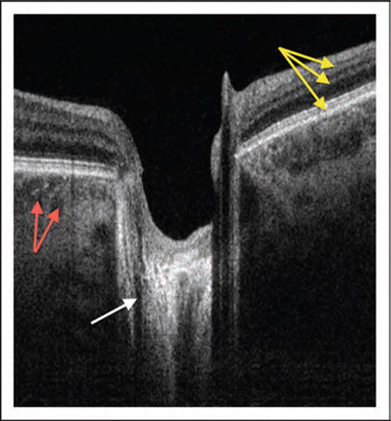
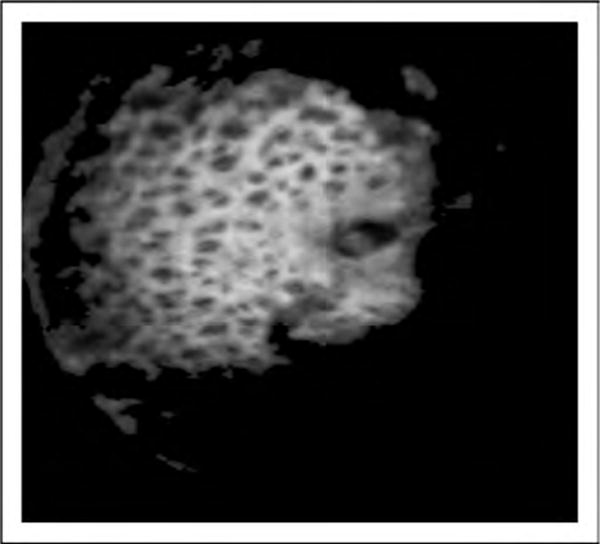
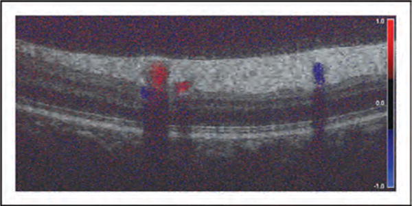
Recent findings: The improvements made in the technology have resulted in increased scanning speed, axial and transverse resolution, and more effective use of the OCT technology as a component of multimodal imaging tools. At the same time, the parallel evolution in novel algorithms makes it possible to efficiently analyze the increased volume of acquired data.
Summary: The innovative iterations of OCT technology have the potential to further improve the performance of the technology in evaluating ocular structural and functional characteristics and longitudinal changes in glaucoma.
Conflict of interest statement
Dr Schuman receives royalties for an optical coherence tomography patent owned and licensed by the Massachusetts Institute of Technology and Massachusetts Eye & Ear Infirmary to Zeiss (Dublin, CA).
Figures
References
-
- Menke MN, Knecht P, Sturm V, et al. Reproducibility of nerve fiber layer thickness measurements using 3D Fourier-domain OCT. Invest Ophthalmol Vis Sci. 2008;49:5386–5391. - PubMed
-
- Francoz M, Fenolland J-R, Giraud J-M, et al. Reproducibility of macular ganglion cell-inner plexiform layer thickness measurement with cirrus HD-OCT in normal, hypertensive and glaucomatous eyes. Br J Ophthalmol. 2014;98:322–328. - PubMed
Publication types
MeSH terms
Grants and funding
LinkOut - more resources
Full Text Sources
Research Materials
Miscellaneous
