Editorial
. 2015 Mar;138(Pt 3):726-35.
doi: 10.1093/brain/awu391. Epub 2015 Jan 15.

Increased sleep need and daytime sleepiness 6 months after traumatic brain injury: a prospective controlled clinical trial

Affiliations
Editorial

Increased sleep need and daytime sleepiness 6 months after traumatic brain injury: a prospective controlled clinical trial

Lukas L Imbach et al. Brain. 2015 Mar.

Abstract

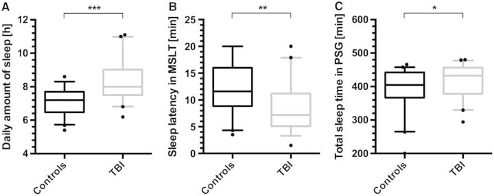
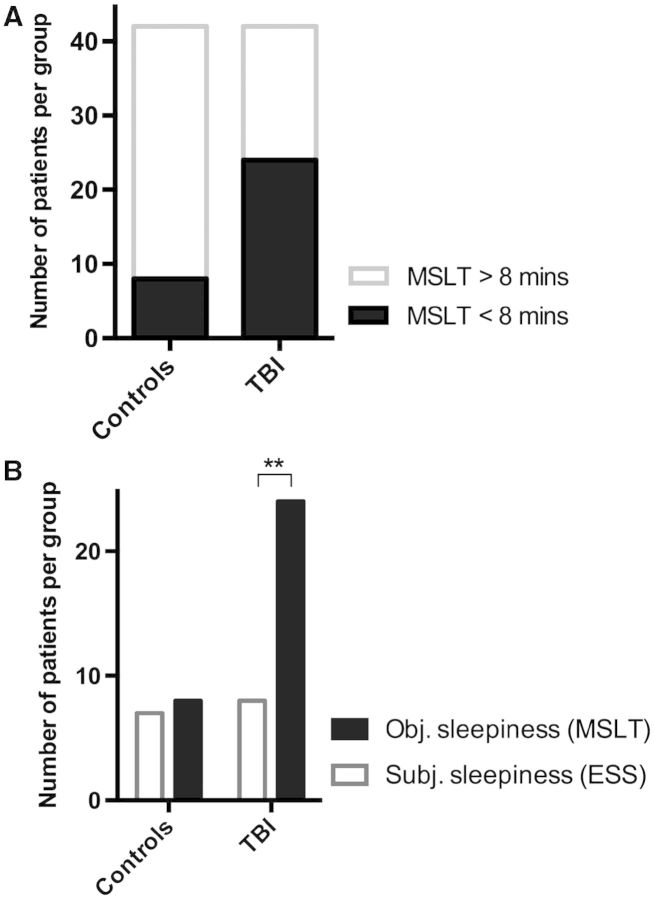
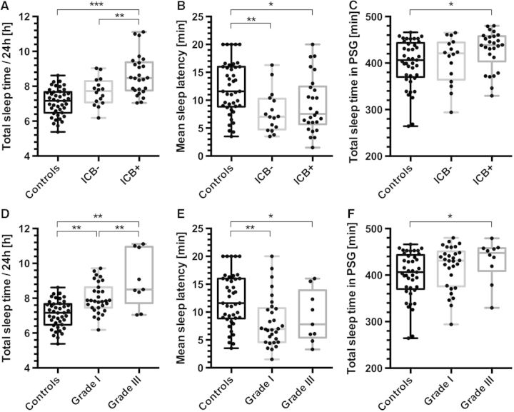
Post-traumatic sleep-wake disturbances are common after acute traumatic brain injury. Increased sleep need per 24 h and excessive daytime sleepiness are among the most prevalent post-traumatic sleep disorders and impair quality of life of trauma patients. Nevertheless, the relation between traumatic brain injury and sleep outcome, but also the link between post-traumatic sleep problems and clinical measures in the acute phase after traumatic brain injury has so far not been addressed in a controlled and prospective approach. We therefore performed a prospective controlled clinical study to examine (i) sleep-wake outcome after traumatic brain injury; and (ii) to screen for clinical and laboratory predictors of poor sleep-wake outcome after acute traumatic brain injury. Forty-two of 60 included patients with first-ever traumatic brain injury were available for follow-up examinations. Six months after trauma, the average sleep need per 24 h as assessed by actigraphy was markedly increased in patients as compared to controls (8.3 ± 1.1 h versus 7.1 ± 0.8 h, P < 0.0001). Objective daytime sleepiness was found in 57% of trauma patients and 19% of healthy subjects, and the average sleep latency in patients was reduced to 8.7 ± 4.6 min (12.1 ± 4.7 min in controls, P = 0.0009). Patients, but not controls, markedly underestimated both excessive sleep need and excessive daytime sleepiness when assessed only by subjective means, emphasizing the unreliability of self-assessment of increased sleep propensity in traumatic brain injury patients. At polysomnography, slow wave sleep after traumatic brain injury was more consolidated. The most important risk factor for developing increased sleep need after traumatic brain injury was the presence of an intracranial haemorrhage. In conclusion, we provide controlled and objective evidence for a direct relation between sleep-wake disturbances and traumatic brain injury, and for clinically significant underestimation of post-traumatic sleep-wake disturbances by trauma patients.

Keywords: post-traumatic daytime sleepiness; post-traumatic pleiosomnia; sleep; traumatic brain injury.


Figures

Figure 1
Figure 1
Overview of study design and patient enrolment. Explanations for loss to follow-up are given in the results section of the manuscript.
Figure 2
Figure 2
Sleep amount and sleep latency in TBI patients and healthy controls. (A) Daily hours of sleep as measured by continuous actigraphy over 2 weeks, showing an increase of 1.2 h in total amount of sleep/24 h in TBI patients as compared to the control group. (B) Mean sleep latencies on the MSLT in TBI patients are 28% lower than in healthy controls. (C) Total sleep time in 8 h-polysomnography (PSG) reveals an increased amount of sleep in TBI patients. Box plots indicate means (horizontal line), upper and lower quartiles (box) and extrema (whiskers), outliers are shown as black dots. ***P < 10−6, **P < 0.0001, *P < 0.05.
Figure 3
Figure 3
Objective versus subjective EDS in TBI patients and controls. (A) Objective EDA (defined as average sleep latency <8 min on MSLT, filled bars) was found in 24/42 TBI patients (57%) as compared to 8/42 healthy subjects (19%; odds ratio: 5.6, P < 0.001). (B) Comparison of subjective EDS (based on Epworth Sleepiness Scale, open bars) and objective EDS (based on MSLT, filled bars). In healthy controls, no difference between subjective and objective measures was found, but in TBI patients, EDS was three times more prevalent when assessed objectively (McNemar objective versus subjective EDS: P > 0.99 in controls and **P < 0.0005 in TBI patients).
Figure 4
Figure 4
Sleep fragmentation in TBI patients and controls. Sleep fragmentation (number of sleep bouts/time) is reduced in NREM sleep in TBI patients (grey bars) as compared to controls (black bars), whereas fragmentation of REM sleep did not differ. Wakefulness was more fragmented in TBI patients (t-test: **P < 0.005, *P < 0.05).
Figure 5
Figure 5
Pleiosomnia and excessive daytime sleepiness with respect to occurrence of intracranial haemorrhage and TBI severity. (A) Patients with intracranial haemorrhage (ICB+) had significantly more sleep per 24 h than both controls and TBI patients without bleedings (ICB−). (B) Mean sleep latencies on MSLT differed between controls and TBI patients, but no significant difference was observed between TBI patients with (ICB+) and without (ICB−) intracranial bleeding (Pearson correlation: R = −0.292, P < 0.01). (C) Patients with intracranial haemorrhage (ICB+) showed increased total sleep time on polysomnography (PSG), whereas sleep time in patients without haemorrhage (ICB−) did not differ from the control group. (D) Patients with severe TBI (Grade III, Glasgow Coma Scale ≤ 8) had more sleep per 24 h than both controls and mild TBI patients (Grade I, Glasgow Coma Scale ≥ 13). (E) Mean sleep latencies on MSLT differed between controls and TBI patients, but not between TBI patients with low and high severity trauma. (F) Patients with Grade III TBI had elevated total sleep time in polysomnography as compared to controls. (***P < 0.001, **P < 0.01 *P < 0.05 in one-way ANOVA, n = 84).
Figure 6
Figure 6
Levels of potential biomarkers in the acute phase versus follow-up after TBI. (A) Acute phase lab values (within 5 days after TBI, grey circles) normalized to values at follow-up (6 months after TBI, dotted line) revealed increased values for TBI markers (S100) and elevated adrenaline levels (*P < 0.05). (B) Change of the diurnal cortisol pattern. The difference between a.m. (black) and p.m. (grey) cortisol levels was diminished in the acute phase (#P = 0.44 for comparison acute versus 6 months, paired two-sided t-test) as compared to 6 months after TBI (##P = 0.06 for comparison acute versus 6 months, unpaired one-sided t-test). (C) Morning cortisol levels (cortisol am) in the acute phase correlated significantly with sleepiness as measured by MSLTs 6 months after TBI (R = 0.88, P = 0.005, n = 10). S100 = S100 calcium binding protein; ACTH = adrenocorticotropic hormone; NSE = neuron-specific enolase.

References

    1. American Academy of Sleep Medicine. . 2nd edn. Westchester, IL: American Academy of Sleep Disorders Association; 2005. International Classification of sleep disorders (ICSD-2), diagnostic and coding manual, 2nd edn.
    1. Baumann CR, Werth E, Stocker R, Ludwig S, Bassetti CL. Sleep-wake disturbances 6 months after traumatic brain injury: a prospective study. Brain. 2007;130:1873–83. - PubMed
    1. Baumann CR, Bassetti CL, Valko PO, Haybaeck J, Keller M, Clark E, et al. Loss of hypocretin (orexin) neurons with traumatic brain injury. Ann Neurol. 2009;66:555–9. - PMC - PubMed
    1. Berger RP, Pierce MC, Wisniewski SR, Adelson PD, Clark RS, Ruppel RA, et al. Neuron-specific enolase and S100B in cerebrospinal fluid after severe traumatic brain injury in infants and children. Pediatrics. 2002;109:E31. - PubMed
    1. Carmichael ST, Chesselet MF. Synchronous neuronal activity is a signal for axonal sprouting after cortical lesions in the adult. J Neurosci. 2002;22:6062–70. - PMC - PubMed

Publication types

MeSH terms