. 2015 Mar;110(2):3.
doi: 10.1007/s00395-014-0459-0. Epub 2015 Jan 17.

Mitochondrially targeted Endonuclease III has a powerful anti-infarct effect in an in vivo rat model of myocardial ischemia/reperfusion

Affiliations

Mitochondrially targeted Endonuclease III has a powerful anti-infarct effect in an in vivo rat model of myocardial ischemia/reperfusion

Xi-Ming Yang et al. Basic Res Cardiol. 2015 Mar.

Abstract

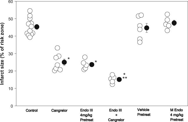
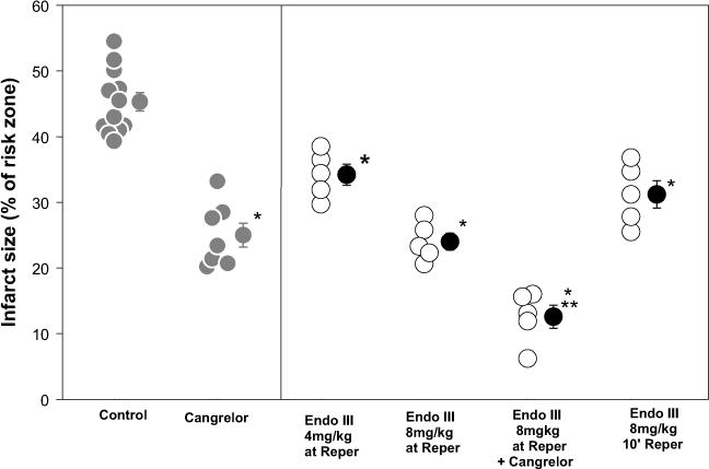
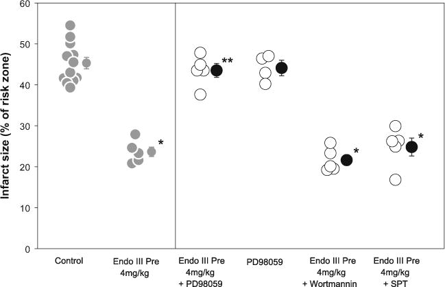
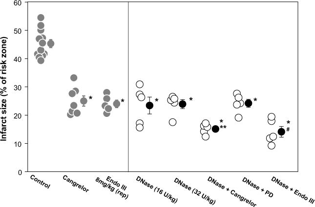
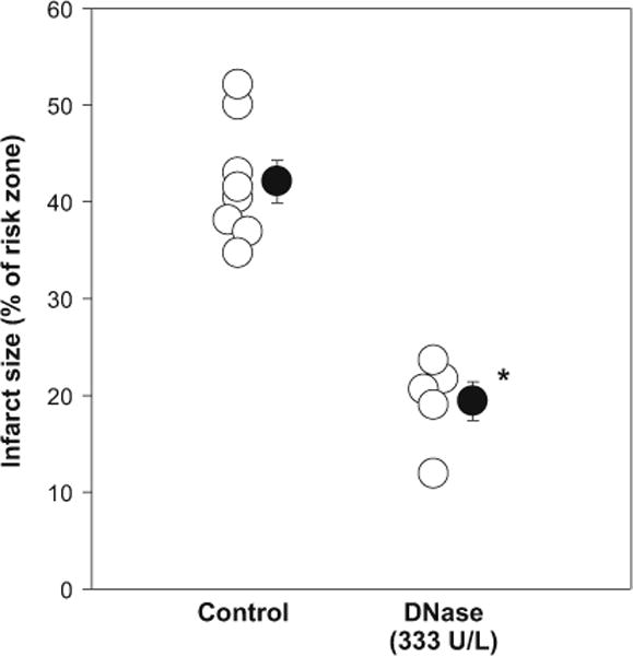
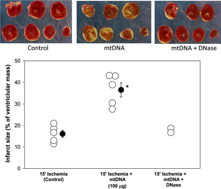
Recent reports indicate that elevating DNA glycosylase/AP lyase repair enzyme activity offers marked cytoprotection in cultured cells and a variety of injury models. In this study, we measured the effect of EndoIII, a fusion protein construct that traffics Endonuclease III, a DNA glycosylase/AP lyase, to the mitochondria, on infarct size in a rat model of myocardial ischemia/reperfusion. Open-chest, anesthetized rats were subjected to 30 min of occlusion of a coronary artery followed by 2 h of reperfusion. An intravenous bolus of EndoIII, 8 mg/kg, just prior to reperfusion reduced infarct size from 43.8 ± 1.4% of the risk zone in control animals to 24.0 ± 1.3% with no detectable hemodynamic effect. Neither EndoIII's vehicle nor an enzymatically inactive EndoIII mutant (K120Q) offered any protection. The magnitude of EndoIII's protection was comparable to that seen with the platelet aggregation inhibitor cangrelor (25.0 ± 1.8% infarction of risk zone). Because loading with a P2Y12 receptor blocker to inhibit platelets is currently the standard of care for treatment of acute myocardial infarction, we tested whether EndoIII could further reduce infarct size in rats treated with a maximally protective dose of cangrelor. The combination reduced infarct size to 15.1 ± 0.9% which was significantly smaller than that seen with either cangrelor or EndoIII alone. Protection from cangrelor but not EndoIII was abrogated by pharmacologic blockade of phosphatidylinositol-3 kinase or adenosine receptors indicating differing cellular mechanisms. We hypothesized that EndoIII protected the heart from spreading necrosis by preventing the release of proinflammatory fragments of mitochondrial DNA (mtDNA) into the heart tissue. In support of this hypothesis, an intravenous bolus at reperfusion of deoxyribonuclease I (DNase I) which should degrade any DNA fragments escaping into the extracellular space was as protective as EndoIII. Furthermore, the combination of EndoIII and DNase I produced additive protection. While EndoIII would maintain mitochondrial integrity in many of the ischemic cardiomyocytes, DNase I would further prevent mtDNA released from those cells that EndoIII could not save from propagating further necrosis. Thus, our mtDNA hypothesis would predict additive protection. Finally to demonstrate the toxicity of mtDNA, isolated hearts were subjected to 15 min of global ischemia. Infarct size doubled when the coronary vasculature was filled with mtDNA fragments during the period of global ischemia. To our knowledge, EndoIII and DNase are the first agents that can both be given at reperfusion and add to the protection of a P2Y12 blocker, and thus should be effective in today's patient with acute myocardial infarction.


Conflict of interest statement

Conflict of interest GW and MNG are co-founders of Exscien, the manufacturer of Endo III.

Figures

Fig. 1
Fig. 1
Infarct size as a % of risk zone in open-chest rats. Open circles represent individual experiments, and closed circles represent group mean ± SEM. Endonuclease III (Endo III) was administered before ischemia (pretreat), while cangrelor infusion was begun 10 min before reperfusion. M Endo is an inactive EndoIII mutant. *p < 0.001 vs Control **p < 0.005 vs Cangrelor and p < 0.01 vs EndoIII Pretreat
Fig. 2
Fig. 2
Infarct size as a % of risk zone in open-chest rats. Open circles represent individual experiments, and closed circles represent group mean ± SEM. Endonuclease III (EndoIII) was administered either 2 min before or 10 min following the onset of reperfusion (Reper), and cangrelor infusion was commenced 10 min before reperfusion. Data points in gray represent groups that have been presented in a previous figure, and are reproduced in this figure to facilitate comparison with new groups. *p < 0.001 vs Control **p < 0.001 vs Cangrelor or EndoIII 4 mg/kg and p < 0.002 vs EndoIII 8 mg/kg at Reper
Fig. 3
Fig. 3
Infarct size as a % of risk zone in open-chest rats. Open circles represent individual experiments, and closed circles represent group mean ± SEM. Endonuclease III (EndoIII) was administered before ischemia (Pre). The signaling antagonist PD98059 or wortmannin or adenosine receptor blocker 8-(sulfophenyl)theophylline (SPT) was infused 5 min before EndoIII. Data points in gray represent groups that have been presented in a previous figure, and are reproduced in this figure to facilitate comparison with new groups. *p < 0.001 vs Control **p < 0.001 vs Endo III Pre
Fig. 4
Fig. 4
Infarct size as a % of risk zone in open-chest rats. Open circles represent individual experiments, and closed circles represent group mean ± SEM. DNase I was infused 5 min before reperfusion (rep) and cangrelor infusion was commenced 10 min before reperfusion. PD98059 (PD) was administered 5 min before DNase I and Endo III (infused 2 min before reperfusion). Data points in gray represent groups that have been presented in a previous figure, and are reproduced in this figure to facilitate comparison with new groups. *p < 0.001 vs Control **p < 0.006 vs Cangrelor and < 0.02 vs DNase 16 and 32 U/kg #p < 0.008 vs DNase 16 and 32 U/kg and p < 0.01 vs Endo III
Fig. 5
Fig. 5
Infarct size as a % of risk zone in isolated, perfused rat hearts with 30 min of regional ischemia. Open circles represent individual experiments, and closed circles represent group mean ± SEM. DNase I was present in the buffer continuously during reperfusion. *p < 0.001 vs Control
Fig. 6
Fig. 6
Infarct size in isolated rat hearts subjected to 15 min of global ischemia and 2 h of reperfusion. Open circles represent individual experiments, and closed circles represent group mean ± SEM. Infarction in slices of representative hearts in each of the three groups is shown. Including purified mitochondrial DNA fragments (mtDNA) in the perfusate at the onset of no-flow ischemia doubled infarct size. Degrading mtDNA with DNase prior to infusion rendered mtDNA innocuous. *p < 0.001 vs. 15′ Ischemia (Control)
Fig. 7
Fig. 7
Western blots of HA-tagged EndoIII in subcellular myocardial compartments and plasma. The left panel shows EndoIII in the total heart homogenate (Total), cytosolic (Cyt), mitochondrial (Mito), and nuclear (Nucl) fractions of normally perfused (N) and ischemic (I) ventricular myocardial samples collected 15 min after reperfusion. EndoIII was injected 5 min prior to reperfusion after 25 min of regional ischemia. Note that EndoIII is concentrated in mitochondria of the ischemic tissue. The right panel shows plasma content of EndoIII just prior to injection of EndoIII (Base), 2 min after injection (2′), and 15 min after reperfusion (15′). Twenty minutes after injection most of the EndoIII had been removed from the blood. The small spot in the Base lane is thought to be contamination from the 2′ lane as all 3 lanes were overloaded in an attempt to see the 15′ signal. ATP synthase is a mitochondria-specific marker. Little mitochondrial contamination is present in the cytosolic fraction but the nuclear fraction shows significant mitochondrial contamination

References

    1. Bliksøen M, Mariero LH, Ohm IK, Haugen F, Yndestad A, Solheim S, Seljeflot I, Ranheim T, Andersen GØ, Aukrust P, Valen G, Vinge LE. Increased circulating mitochondrial DNA after myocardial infarction. Int J Cardiol. 2012;158:132–134. doi: 10.1016/j.ijcard.2012.04.047. - DOI - PubMed
    1. Boiteux S, Guillet M. Abasic sites in DNA: repair and biological consequences in Saccharomyces cerevisiae. DNA Repair. 2004;3:1–12. doi: 10.1016/j.dnarep.2003.10.002. - DOI - PubMed
    1. Burney S, Caulfield JL, Niles JC, Wishnok JS, Tannenbaum SR. The chemistry of DNA damage from nitric oxide and peroxynitrite. Mutat Res. 1999;424:37–49. doi: 10.1016/S0027-5107(99)00006-8. - DOI - PubMed
    1. Chatterjee A, Mambo E, Zhang Y, DeWeese T, Sidransky D. Targeting of mutant hogg1 in mammalian mitochondria and nucleus: effect on cellular survival upon oxidative stress. BMC Cancer. 2006;6:235. doi: 10.1186/1471-2407-6-235. - DOI - PMC - PubMed
    1. Chouteau JM, Obiako B, Gorodnya OM, Pastukh VM, Ruchko MV, Wright AJ, Wilson GL, Gillespie MN. Mitochondrial DNA integrity may be a determinant of endothelial barrier properties in oxidant-challenged rat lungs. Am J Physiol. 2011;301:L892–L898. doi: 10.1152/ajplung.00210.2011. - DOI - PMC - PubMed

Publication types

MeSH terms

LinkOut - more resources