. 2015 Mar;94(3):439-45.
doi: 10.1177/0022034514567198. Epub 2015 Jan 16.

Wntless regulates dentin apposition and root elongation in the mandibular molar

Affiliations

Wntless regulates dentin apposition and root elongation in the mandibular molar

C H Bae et al. J Dent Res. 2015 Mar.

Abstract

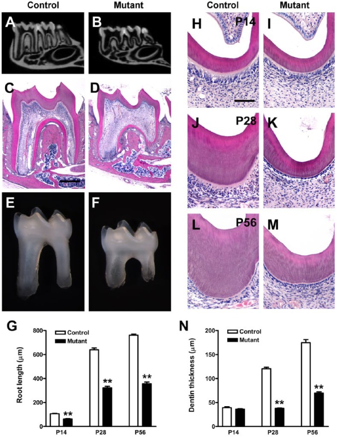
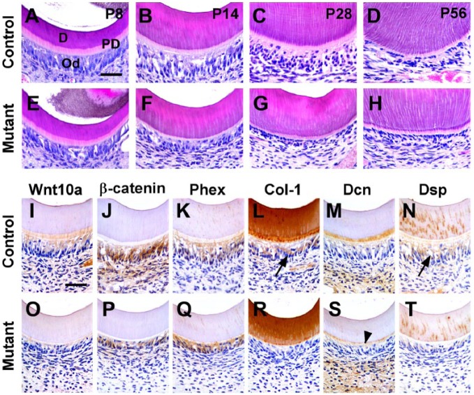
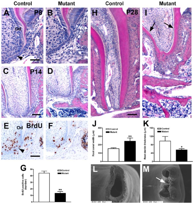
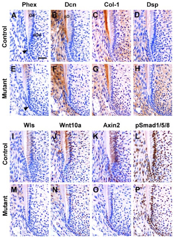
Wnt signaling plays an essential role in the dental epithelium and mesenchyme during tooth morphogenesis. However, it remains unclear if Wnt ligands, produced from dental mesenchyme, are necessary for odontoblast differentiation and dentin formation. Here, we show that odontoblast-specific disruption of Wntless (Wls), a chaperon protein that regulates Wnt sorting and secretion, leads to severe defects in dentin formation and root elongation. Dentin thickness decreased remarkably and pulp chambers enlarged in the mandibular molars of OC-Cre;Wls(CO/CO) mice. Although the initial odontoblast differentiation was normal in the mutant crown, odontoblasts became cuboidal and dentin thickness was reduced. In immunohistochemistry, Wnt10a, β-catenin, type I collagen, and dentin sialoprotein were significantly down-regulated in the odontoblasts of mutant crown. In addition, roots were short and root canals were widened. Cell proliferation was reduced in the developing root apex of mutant molars. Furthermore, Wnt10a and Axin2 expression was remarkably decreased in the odontoblasts of mutant roots. Deletion of the Wls gene in odontoblasts appears to reduce canonical Wnt activity, leading to inhibition of odontoblast maturation and root elongation.

Keywords: Wnt signaling pathway; dentinogenesis; mice; odontoblasts; tooth development; tooth roots.


Conflict of interest statement

The authors declare no potential conflicts of interest with respect to the authorship and/or publication of this article.

Figures

Figure 1.
Figure 1.
Tooth phenotypes in OC-Cre;WlsCO/CO mice. Mandibular molars of mutant mice exhibited thin dentin, enlarged pulp chambers, and short roots. (A, B) Micro–computed tomographic view of mandibles from control and mutant mice at P28. (C–F) Histologic features and stereoscopic appearance of the mandibular first molar of control and mutant mice at P28. (G) Quantification of root length of the mandibular first molar at P14, P28, and P56 (n = 6, in each genotype for stages, respectively). (H–M) Hematoxylin and eosin–stained sections of crown dentin at P14, P28, and P56. (N) Differences in dentin thickness of controls and mutants at P14, P28, and P56 (n = 6, in each genotype for stages, respectively). **P < 0.01. Scale bars: 400 µm (C, D), 100 µm (H–M).
Figure 2.
Figure 2.
Morphologic and molecular changes in crown odontoblasts of OC-Cre;WlsCO/CO mice during dentin formation. (A–H) Histologic features of odontoblasts in the crowns of control and mutant mice at P8, P14, P28, and P56. (I–T) Localization of Wnt10a, β-catenin, Phex, Col-1, Dcn, and Dsp on the crown odontoblasts of the mandibular first molar at P14. Black arrowhead indicates predentin. D, dentin; Od, odontoblasts; PD, predentin. Scale bars: 40 µm (A–H), 100 µm (I–T).
Figure 3.
Figure 3.
Impaired root elongation in the mandibular first molar of OC-Cre;WlsCO/CO mice. (A–D, H–I) Hematoxylin and eosin–stained sections of the mandibular first molar in control and mutant mice at P8, P14, and P28. (E–G) In the developing root apex of controls at P14, BrdU-labeled proliferating cells were abundant. However, few cells were positively labeled with BrdU in mutant mice. (J–K) Root canal width and radicular dentin thickness in control and mutant mice at P28. (L–M) Basal view of distal root apex of the mandibular first molar at P28. *P < 0.05 and **P < 0.01. Black arrowhead indicates Hertwig’s epithelial root sheath. Od, odontoblasts. Scale bars: 40 µm (A, B), 80 µm (E, F), and 100 µm (C, D, G, H).
Figure 4.
Figure 4.
Inhibition of odontoblast maturation during root elongation in developing molar roots of OC-Cre;WlsCO/CO mice. (A, E) Both in controls and mutants, Phex was localized in mature odontoblasts but not in differentiating odontoblasts. (B, F) In controls, Dcn specifically marked predentin, which was thinner in mutants. (C, D, G, H) Col-1 and Dsp were localized in dentin in both controls and mutants. (I, M) Wls was localized in mature odontoblasts and differentiating odontoblasts in control but was not found in mutant odontoblasts. (J, K, N, O) Wnt10a and Axin2 were widely expressed in control odontoblasts, differentiating odontoblasts, and Hertwig’s epithelial root sheath (HERS) cells, but their overall expression was decreased in mutants. (L, P) pSmad1/5/8 was abundantly expressed in both control and mutant odontoblasts, HERS, and pulp cells. Black arrowhead indicates differentiating odontoblasts. Od, odontoblasts; dOd, differentiating odontoblasts; PD, predentin. Scale bars: 40 µm (A–P).

References

    1. Banziger C, Soldini D, Schütt C, Zipperlen P, Hausmann G, Basler K. 2006. Wntless, a conserved membrane protein dedicated to the secretion of Wnt proteins from signaling cells. Cell. 125(3):509–522. - PubMed
    1. Bartscherer K, Pelte N, Ingelfinger D, Boutros M. 2006. Secretion of Wnt ligands requires Evi, a conserved transmembrane protein. Cell. 125(3):523–533. - PubMed
    1. Carpenter AC, Rao S, Wells JM, Campbell K, Lang RA. 2010. Generation of mice with a conditional null allele for Wntless. Genesis. 48(9):554–558. - PMC - PubMed
    1. Chen J, Lan Y, Baek JA, Gao Y, Jiang R. 2009. Wnt/beta-catenin signaling plays an essential role in activation of odontogenic mesenchyme during early tooth development. Dev Biol. 334(1):174–185. - PMC - PubMed
    1. Fjeld K, Kettunen P, Furmanek T, Kvinnsland IH, Luukko K. 2005. Dynamic expression of Wnt signaling-related Dickkopf1, -2, and -3 mRNAs in the developing mouse tooth. Dev Dyn. 233(1):161–166. - PubMed

Publication types

MeSH terms

LinkOut - more resources