. 2015 Mar;81(6):2233-43.
doi: 10.1128/AEM.03733-14. Epub 2015 Jan 16.

Elizabethkingia anophelis: molecular manipulation and interactions with mosquito hosts

Affiliations

Elizabethkingia anophelis: molecular manipulation and interactions with mosquito hosts

Shicheng Chen et al. Appl Environ Microbiol. 2015 Mar.

Abstract

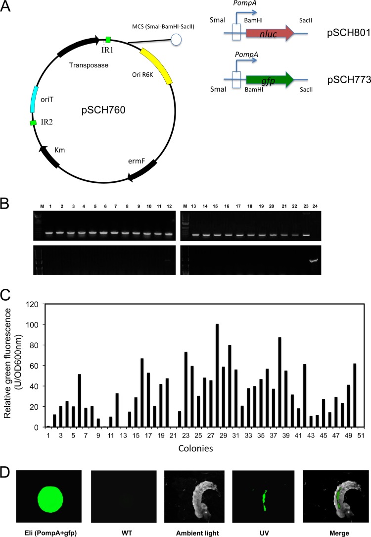
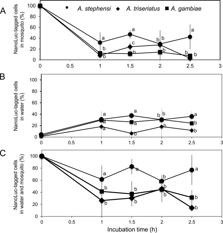
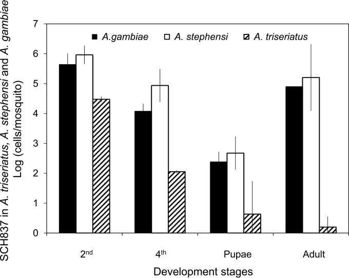
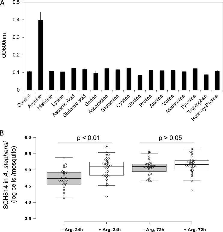
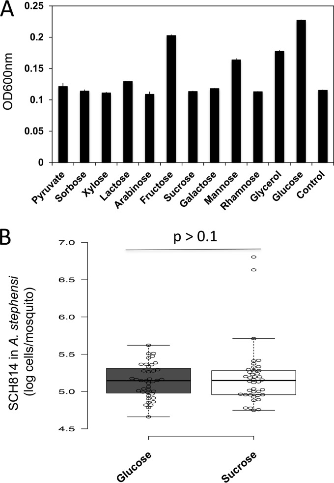
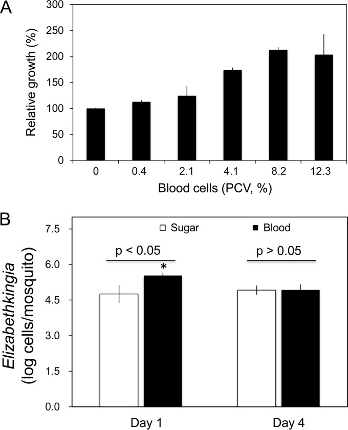
Flavobacteria (members of the family Flavobacteriaceae) dominate the bacterial community in the Anopheles mosquito midgut. One such commensal, Elizabethkingia anophelis, is closely associated with Anopheles mosquitoes through transstadial persistence (i.e., from one life stage to the next); these and other properties favor its development for paratransgenic applications in control of malaria parasite transmission. However, the physiological requirements of E. anophelis have not been investigated, nor has its capacity to perpetuate despite digestion pressure in the gut been quantified. To this end, we first developed techniques for genetic manipulation of E. anophelis, including selectable markers, reporter systems (green fluorescent protein [GFP] and NanoLuc), and transposons that function in E. anophelis. A flavobacterial expression system based on the promoter PompA was integrated into the E. anophelis chromosome and showed strong promoter activity to drive GFP and NanoLuc reporter production. Introduced, GFP-tagged E. anophelis associated with mosquitoes at successive developmental stages and propagated in Anopheles gambiae and Anopheles stephensi but not in Aedes triseriatus mosquitoes. Feeding NanoLuc-tagged cells to A. gambiae and A. stephensi in the larval stage led to infection rates of 71% and 82%, respectively. In contrast, a very low infection rate (3%) was detected in Aedes triseriatus mosquitoes under the same conditions. Of the initial E. anophelis cells provided to larvae, 23%, 71%, and 85% were digested in A. stephensi, A. gambiae, and Aedes triseriatus, respectively, demonstrating that E. anophelis adapted to various mosquito midgut environments differently. Bacterial cell growth increased up to 3-fold when arginine was supplemented in the defined medium. Furthermore, the number of NanoLuc-tagged cells in A. stephensi significantly increased when arginine was added to a sugar diet, showing it to be an important amino acid for E. anophelis. Animal erythrocytes promoted E. anophelis growth in vivo and in vitro, indicating that this bacterium could obtain nutrients by participating in erythrocyte lysis in the mosquito midgut.


Figures

FIG 1
FIG 1
Reporter strain construction and demonstration of E. anophelis cells tagged with GFP and their ingestion by mosquito larvae. (A) Diagram of the pSCH760 construct. pHimarEm1(MCS) was modified with a multiple-cloning site (SmaI-BamHI-SacII); the expression cassettes PompA+gfp or PompA+nluc were inserted into SmaI and SacII sites on pSCH760 to generate pSCH773 and pSCH801, respectively. (B) The transposon incorporated into the E. anophelis MSU001 chromosome. (Upper panel) PCR screening results for the Em-resistant transconjugants, using the primers Walker140 and Walker141. Lane M, molecular marker; lanes 1 to 22, DNA fragments amplified from Em-resistant transconjugants; lane 23, positive control (pSCH760 as the amplification template); lane 24, negative control (E. anophelis MSU001). (Lower panel) Presence of transposase, determined using primers Walker186 and Walker187. Lanes 1 to 22, the same transconjugants as in the upper panel; lane 23, negative control (E. anophelis MSU001); lane 24, positive control (pSCH760 as the amplification template). (C) Quantitative analysis of E. anophelis emitting GFP fluorescence. Transconjugants were first screened under UV, and the fluorescent colonies were next quantified using fluorometry. The brightest colony was chosen for further study. (D) The cultures carrying the GFP reporter were incubated with Anopheles mosquito larvae for 2 h, and the larvae were observed by using epifluorescence microscopy. The control was E. anophelis MSU001 cells.
FIG 2
FIG 2
Digestibility analysis of E. anophelis by Aedes triseriatus, A. gambiae, and A. stephensi larvae. (A) Larval mosquitoes fed SCH814 cells were pooled (4 at each time point), homogenized, washed, and subjected to the NanoLuc activity assay. Cell densities at the different time points were normalized to the initial cell densities in corresponding mosquitoes at time zero. (B) Cells in the water were sampled, washed with PBS by centrifuging, resuspended in PBS, and subjected to the NanoLuc activity assay. Cell densities at the different time points were normalized to the initial cell densities at time zero. (C) The NanoLuc-tagged cells recovered from mosquitoes and water samples were quantified and normalized to those at time zero. Values are means ± standard deviations; triplicate experiments were performed. Significant differences among Aedes triseriatus, A. gambiae, and A. stephensi samples at each time point were determined by using PROC GLM. Different letters (a, b, and c) indicate significant differences in NanoLuc-tagged cell densities among these samples at each time point (P < 0.05). Means with the same letters indicate that no statistically significant difference was observed for these samples (P > 0.05).
FIG 3
FIG 3
Association of introduced E. anophelis with mosquitoes. Cells tagged with GFP were introduced to A. gambiae, A. stephensi, and Aedes triseriatus second-instar mosquito larvae. The numbers of CFU were counted and calculated by plating homogenized mosquito samples (pools of 5 mosquitoes) on LB plates containing Em. Values are means ± standard deviations; triplicate experiments were performed.
FIG 4
FIG 4
Effects of amino acids on SCH814 growth in vivo and in vitro. (A) Selected amino acids at 4 mM (final concentration) were individually added to M9 medium with glucose. After 24 h of incubation at 30°C, the cells were subjected to optical density determinations at 600 nm. (B) A. stephensi mosquitoes were fed 10% sucrose supplemented with SCH814 for 24 h (NanoLuc reporter strain). After the adult mosquitoes emerged, they were fed 10% sucrose with 10 mM arginine or 10% sucrose without arginine. After 24 h and 72 h, 30 mosquitoes were randomly sampled from sucrose without arginine treatment at each time point. Under the same conditions, 28 and 30 mosquitoes were sampled from sucrose with 10 mM arginine treatment, respectively. Mosquitoes were homogenized and subjected to NanoLuc assays. Significant differences between arginine addition and no-arginine addition samples were determined by using PROC GLM; significantly different cell densities are denoted by an asterisk (P < 0.05).
FIG 5
FIG 5
Effects of carbon source on SCH814 growth in vivo and in vitro. (A) Selected carbon sources at a 0.5% (wt/vol) final concentration were added to M9 medium. After 24 h of incubation at 30°C, the cells were subjected to optical density determinations at 600 nm. (B) Second-instar larvae (A. stephensi) were inoculated with SCH814 (NanoLuc reporter strain). After the adult mosquitoes emerged, they were fed 10% glucose or 10% sucrose. Thirty mosquitoes from each treatment group were randomly sampled, homogenized, and subjected to NanoLuc assays. Significant differences between glucose and sucrose samples were determined by using PROC GLM.
FIG 6
FIG 6
Effects of animal blood on SCH814 growth in vivo and in vitro. (A) Effect of different concentrations of animal blood cells on SCH814 growth in M9 medium in vitro. SCH814 cells were cultured in M9 medium (with glucose) supplemented with various concentrations of horse blood cells. After incubation at 30°C for 24 h, SCH814 cells were estimated by the determination of NanoLuc activity; the relative growth was expressed as the percentage relative to the control (without supplementation with horse blood cells, set as 100%). Values are means of single measurements from triplicate experiments (± standard deviations). (B) Effect of sugar and blood meals on SCH814 growth in mosquitoes. A suspension of SCH814 cells in 10% sucrose was fed to A. stephensi mosquitoes for 24 h in order to introduce NanoLuc-tagged bacteria. The mosquitoes were then given sugar meal (10% sucrose) or blood meal via a membrane apparatus (see Materials and Methods). Four mosquitoes from each treatment group were sampled for assay of NanoLuc activity on day 1. Under the same testing conditions, 8 mosquitoes from each treatment group were sampled on day 4. Significant differences between sugar and blood meals were determined by using PROC GLM and are denoted by an asterisk (P < 0.05).

References

    1. Wang Y, Gilbreath TM, Kukutla P, Yan G, Xu J. 2011. Dynamic gut microbiome across life history of the malaria mosquito Anopheles gambiae in Kenya. PLoS One 6:e24767. doi:10.1371/journal.pone.0024767. - DOI - PMC - PubMed
    1. Kukutla P, Lindberg BG, Pei D, Rayl M, Yu W, Steritz M, Faye I, Xu J. 2014. Insights from the genome annotation of Elizabethkingia anophelis from the malaria vector Anopheles gambiae. PLoS One 9:e97715. doi:10.1371/journal.pone.0097715. - DOI - PMC - PubMed
    1. Boissière A, Tchioffo MT, Bachar D, Abate L, Marie A, Nsango SE, Shahbazkia HR, Awono-Ambene PH, Levashina EA, Christen R, Morlais I. 2012. Midgut microbiota of the malaria mosquito vector Anopheles gambiae and interactions with Plasmodium falciparum infection. PLoS Pathog 8:e1002742. doi:10.1371/journal.ppat.1002742. - DOI - PMC - PubMed
    1. Terenius O, Lindh JM, Eriksson-Gonzales K, Bussière L, Laugen AT, Bergquist H, Titanji K, Faye I. 2012. Midgut bacterial dynamics in Aedes aegypti. FEMS Microbiol Ecol 80:556–565. doi:10.1111/j.1574-6941.2012.01317.x. - DOI - PubMed
    1. Akhouayri IG, Habtewold T, Christophides GK. 2013. Melanotic pathology and vertical transmission of the gut commensal Elizabethkingia meningoseptica in the major malaria vector Anopheles gambiae. PLoS One 8:e77619. doi:10.1371/journal.pone.0077619. - DOI - PMC - PubMed

Publication types

Substances

Associated data

LinkOut - more resources