Review
. 2015:129:285-326.
doi: 10.1016/bs.pmbts.2014.10.012. Epub 2014 Dec 1.

Molecular determinants of the ratio of inert to infectious virus particles

Affiliations
Review

Molecular determinants of the ratio of inert to infectious virus particles

P J Klasse. Prog Mol Biol Transl Sci. 2015.

Abstract

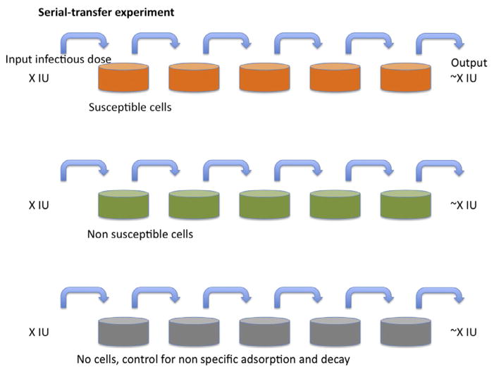
The ratio of virus particles to infectious units is a classic measurement in virology and ranges widely from several million to below 10 for different viruses. Much evidence suggests a distinction be made between infectious and infecting particles or virions: out of many potentially infectious virions, few infect under regular experimental conditions, largely because of diffusion barriers. Still, some virions are inert from the start; others become defective through decay. And with increasing cell- and molecular-biological knowledge of each step in the replicative cycle for different viruses, it emerges that many processes entail considerable losses of potential viral infectivity. Furthermore, all-or-nothing assumptions about virion infectivity are flawed and should be replaced by descriptions that allow for spectra of infectious propensities. A more realistic understanding of the infectivity of individual virions has both practical and theoretical implications for virus neutralization, vaccine research, antiviral therapy, and the use of viral vectors.

Keywords: Attachment; Defective particles; Endocytosis; Entry; Fusion; Gene therapy; HIV; Infectious unit; Restriction; Transcription; Uncoating; Virion; Viruses.


Figures

Figure 1
Figure 1. Serial transfer
A nominal infectious dose (X IU) of virus is added to the tissue-culture dishes in the left-hand column. After incubation for a fixed number of hours and at a constant temperature, the medium is aspirated from the second dish, to which all of the medium is transferred from the first dish, which is replenished with medium without virus. The procedure is repeated until the right-most dish has been incubated with supernatant for the same time as the others. The top row of dishes (orange (gray in the print version)) have susceptible cells forming a nonconfluent cell matt. The middle row (green (gray in the print version)) have nonsusceptible but otherwise similar cells (for example, lacking only a crucial receptor) growing at the same confluency. The bottom row has only tissue-culture dishes without cells. The outcome of the classical experiment as outlined is usually that infectious virus is transferred from the left to the right at nearly constant levels as detected in each dish; or there may be nonspecific losses, for example, some virus is lost for each step also in the middle row by binding to cell-surface glycosaminoglycans, equally abundant on nonsusceptible cells; or the non-specific losses through binding only to plastic (bottom row) may be equally great; or the infectivity declines significantly in the medium within the time-frame of the experiment (not shown). The key finding is that the losses attributable to infection are negligible and that is because the diffusion time from the top to the bottom of the medium in the tissue-culture dish is too long for more than a small fraction of the virions to encounter target cells through diffusion.
Figure 2
Figure 2. Volume versus concentration
Four tissue-culture wells are shown: the first, third, and fourth from the left are given the same amount of input virus; the second is given twice as much. In the two tissue-culture wells to the left, medium containing the same concentration of infectious virions is added. In the third one, the concentration is half of that for the two to the left but in the fourth it is double that (the volume is half of that of first one to the left). Experiments of this sort tend to show that the infectivity readout in the two wells to the left is similar; it is twice as low in the third one and twice as high in the fourth. Measured P/IU would have varied accordingly although the virus preparation was the same and hence the real P/IU was constant, as well as lower than all those based on measurements determined by the slow diffusion.
Figure 3
Figure 3. Infectivity decay in suspension and abortive events during attachment and after entry
While virions are diffusing at random, they may lose their infectivity and become inert particles (gray) because of the exponential decay of their components; for enveloped viruses the decay of the envelope glycoproteins may often determine the infectivity half-life. The virion (red) is depicted as getting endocytosed (the endosomal lumen is bright green) and as it later either fuses with or penetrates from the endosome. In case of excessive delay before cytoplasmic entry, the endosome continues to the lysosomal compartment and the virion gets degraded (gray). In case of successful entry of the capsid into the cytoplasm, a productive uncoating (orange) may lead to genome (green curved double lines) release for transcription or, in competition with this, degradation of the whole core by the proteasome (yellow flash).

References

    1. Bhattacharya B, Weiss RA, Davis C, Holmes H, Hockley D, Fassati A. Detection and quantitation of human immunodeficiency virus type-1 particles by confocal microscopy. J Virol Methods. 2004;120:13–21. - PubMed
    1. Heider S, Metzner C. Quantitative real-time single particle analysis of virions. Virology. 2014;462–463C:199–206. - PMC - PubMed
    1. Kimpton J, Emerman M. Detection of replication-competent and pseudotyped human immunodeficiency virus with a sensitive cell line on the basis of activation of an integrated beta-galactosidase gene. J Virol. 1992;66:2232–2239. - PMC - PubMed
    1. Klasse PJ, McKeating JA. Soluble CD4 and CD4 immunoglobulin-selected HIV-1 variants: a phenotypic characterization. AIDS Res Hum Retroviruses. 1993;9:595–604. - PubMed
    1. Flint SJ, Enquist LW, Racaniello VR, Skalka AM. Principles of Virology: Molecular Biology, Pathogenesis, and Control of Animal Viruses. 2. Washington, DC: ASM Press; 2004.

Publication types