. 2015 Jan 30;6(3):1396-413.
doi: 10.18632/oncotarget.2751.

Amyloid accumulation is a late event in sporadic Alzheimer's disease-like pathology in nontransgenic rats

Affiliations

Amyloid accumulation is a late event in sporadic Alzheimer's disease-like pathology in nontransgenic rats

Natalia A Stefanova et al. Oncotarget. .

Abstract

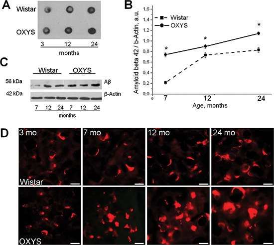
The amyloid cascade hypothesis posits that deposition of the amyloid β (Aβ) peptide in the brain is a key event in the initiation of Alzheimer's disease (AD). Nonetheless, it now seems increasingly unlikely that amyloid toxicity is the cause of sporadic AD, which leads to cognitive decline. Here, using accelerated-senescence nontransgenic OXYS rats, we confirmed that aggregation of Aβ is a later event in AD-like pathology. We showed that an age-dependent increase in the levels of Aβ₁₋₄₂ and extracellular Aβ deposits in the brain of OXYS rats occur later than do synaptic losses, neuronal cell death, mitochondrial structural abnormalities, and hyperphosphorylation of the tau protein. We identified the variants of the genes that are strongly associated with the risk of either late-onset or early-onset AD, including App, Apoe4, Bace1, Psen1, Psen2, and Picalm. We found that in OXYS rats nonsynonymous SNPs were located only in the genes Casp3 and Sorl1. Thus, we present proof that OXYS rats may be a model of sporadic AD. It is possible that multiple age-associated pathological processes may precede the toxic amyloid accumulation, which in turn triggers the final stage of the sporadic form of AD and becomes a hallmark event of the disease.


Conflict of interest statement

Conflict of interest

The authors declare that they have no competing financial interests.

Figures

Figure 1
Figure 1. Deposition of Aβ variants in the brain of OXYS rats
(A, B, E–H), and (J) Brain slices were immunostained for Aβ, with an Aβ1–42- and MOAB-2-specific antibody, respectively. Panel (C) shows the Kongo Red-stained plaques in the cortex, and the same plaques were identified by counterstaining with Sirius Red (D). Photomicrographs (I, J) and (K) demonstrate that the fibrillar Aβ deposits can be detected by Thioflavin-S. Panel (M) shows vascular Aβ deposits identified by counterstaining with Thioflavin-S. The scale bar is 50 μm in (E, F), and (G); 20 μm in (A, C), and (H–J); and 10 μm in (B, D, K), and (L).
Figure 2
Figure 2. The pronounced accumulation of Aβ1–42 with age in the brain of OXYS rats
(A) Dot-blot analysis of Aβ1–42 probed with the MOAB-2 antibody in the hippocampus of 3-, 12-, and 24-month-old OXYS and Wistar rats. Equal amounts of total protein were loaded on each dot. Aβ1–42 indeed accumulates more strongly by the age of 12 and 24 months in the brain of OXYS rats, but there were no differences in the MOAB-2 immunoreactivity between the two strains at 3 months of age. (B–C) Western blot analysis of TBS-soluble oligomeric Aβ1–42 probed with the specific antibody that detects Aβ1–42, and does not detect Aβ1–40, full-length APP, sAPP beta or sAPP alpha. The increased levels of Aβ1–42 detected according to quantitative in 7-, 12-, and 24-month-old OXYS rats compared to Wistar rats. Legend: *statistically significant differences between the strains of the same age. (D) Photomicrographs demonstrate the intensive aggregation of amyloid in the hippocampus of 7-, 12-, and 24- month-old OXYS rats but not at the age of 3 months, according to immunoreactivity with Aβ1–42-specific antibodies. The scale bar is 10 μm.
Figure 3
Figure 3. Increased expression of tau and the increased proportion of insoluble tau in OXYS rats. Expression of tau was analyzed by western blotting in the hippocampus of 3-, 12-, and 24-month-old OXYS and Wistar rats. The data are presented as a percentage of the data from 3-month-old Wistar rats in a group
(A, C) The level of total tau (normalized to the level of β-actin) was significantly increased in OXYS rats compared to Wistar rats in each age group. (B, D) The proportion of insoluble tau and the ratio of sarkosyl-insoluble tau to sarkosyl-soluble tau (normalized to the level of GAPDH and β-actin, respectively) were significantly increased in the hippocampus of 3-, 12-, and 24-month-old OXYS rats compared to Wistar rats. The results were normalized to total protein and are presented as mean ± SEM of at least five independent experiments performed in duplicate. Legend: *statistically significant differences between the strains of the same age; #significant differences with the previous age within the strain.
Figure 4
Figure 4. Increased phosphorylation of sarkosyl-insoluble tau in OXYS rats
Expression of the sarkosyl-soluble fraction and sarkosyl-insoluble fraction recognized by the T181 antibodies was analyzed by western blotting in the hippocampus of 3-, 12-, and 24-month-old OXYS and Wistar rats. The data are presented as a percentage of the data from 3-month-old Wistar rats in a group. (A, C) The level of the sarkosyl-soluble fraction (normalized to β-actin) significantly decreased in OXYS rats only at 3 months of age compared to Wistar rats. (B, C) The level of the sarkosyl-insoluble fraction (normalized to GAPDH) significantly increased in the hippocampus of 3-, 12-, and 24-month-old OXYS rats compared to Wistar rats. (D) The proportion of insoluble phosphotau and the ratio of sarkosyl-insoluble phosphotau to sarkosyl-soluble phosphotau significantly increased in the hippocampus of 3-, 12-, and 24-month-old OXYS rats compared to the Wistar strain. The presented results were normalized to total protein and represent mean ± SEM of at least five independent experiments performed in duplicate. Legend: *statistically significant differences between the strains of the same age; #significant differences compared to the previous age within the strain.
Figure 5
Figure 5. Alterations in the ultrastructure of the CA1 pyramidal neurons of OXYS rats
(A) Mitochondria with normal ultrastructure in the cytoplasm of a neuron from a 4-month-old Wistar rat. (B) Mitochondria with a light matrix in the cytoplasm of a neuron of a 4-month-old OXYS rat. (C) Mitochondria with enlarged and empty intermembrane space in an 18-month-old Wistar rat. (D) Degradation of mitochondria in the neuronal cytoplasm of an 18-month-old OXYS rat. (E) The area occupied by mitochondria within the pyramidal neurons of the hippocampus as % of the total neuron area in the Wistar and OXYS rats. Legend: *statistically significant differences between the strains of the same age.
Figure 6
Figure 6. Synaptic losses and progressive neurodegeneration in OXYS rats
(A) Immunohistochemical analysis using antibodies against PSD-95 (green) and synapsin I (Syn 1; red) on cortex neurons of 18-month-old OXYS and Wistar rats. The scale bar is 5 μm. (B, C) Quantification of the density of PSD-95 and synapsin I in the CA1 region (B) and in the cortex (C) of 4- and 18-month-old OXYS and Wistar rats. The data are presented as a percentage of the data from 4-month-old Wistar rats in a group. (D, E) Expression of PSD-95 and synapsin I was analyzed by western blotting in the hippocampus (D) and in the cortex (E) of 3-, 12-, and 24-month-old OXYS and Wistar rats. (F) Quantification of structural changes of hippocampal and cortical neurons of 4- and 18-month-old OXYS and Wistar rats (as a percentage of dead or damaged neurons). (G) Photomicrographs show the cortex neurons of 4-month-old Wistar rats and 4- and 18-month-old OXYS rats. The arrow shows damaged neurons in OXYS rats. The neurons were stained with Cresyl Violet. (B–E) Values are mean ± SEM. Legend: *statistically significant differences between the strains of the same age; #significant differences with the previous age within the strain.

References

    1. Morley JE, Armbrecht HJ, Farr SA, Kumar VB. The senescence accelerated mouse (SAMP8) as a model for oxidative stress and Alzheimer's disease. Biochim Biophys Acta. 2012;1822:650–656. - PubMed
    1. Drachman DA. The amyloid hypothesis, time to move on: Amyloid is the downstream result, not cause, of Alzheimer's disease. Alzheimers Dement. 2014;3:372–380. - PubMed
    1. Morley JE, Farr SA. The role of amyloid-beta in the regulation of memory. Biochem Pharmacol. 2014;88:479–485. - PubMed
    1. Krstic D, Knuesel I. Deciphering the mechanism underlying late-onset Alzheimer disease. Nat Rev Neurol. 2013;9:25–34. - PubMed
    1. Castellano JM, Deane R, Gottesdiener AJ, Verghese PB, Stewart FR, West T, Paoletti AC, Kasper TR, DeMattos RB, Zlokovic BV, Holtzman DM. Low-density lipoprotein receptor overexpression enhances the rate of brain-to-blood Aβ clearance in a mouse model of β-amyloidosis. Proc Natl Acad Sci U S A. 2012;109:15502–15507. - PMC - PubMed

Publication types

Substances