. 2015 May 1;24(9):2458-69.
doi: 10.1093/hmg/ddv007. Epub 2015 Jan 16.

Crystal structure of human persulfide dioxygenase: structural basis of ethylmalonic encephalopathy

Affiliations

Crystal structure of human persulfide dioxygenase: structural basis of ethylmalonic encephalopathy

Ilaria Pettinati et al. Hum Mol Genet. .

Abstract

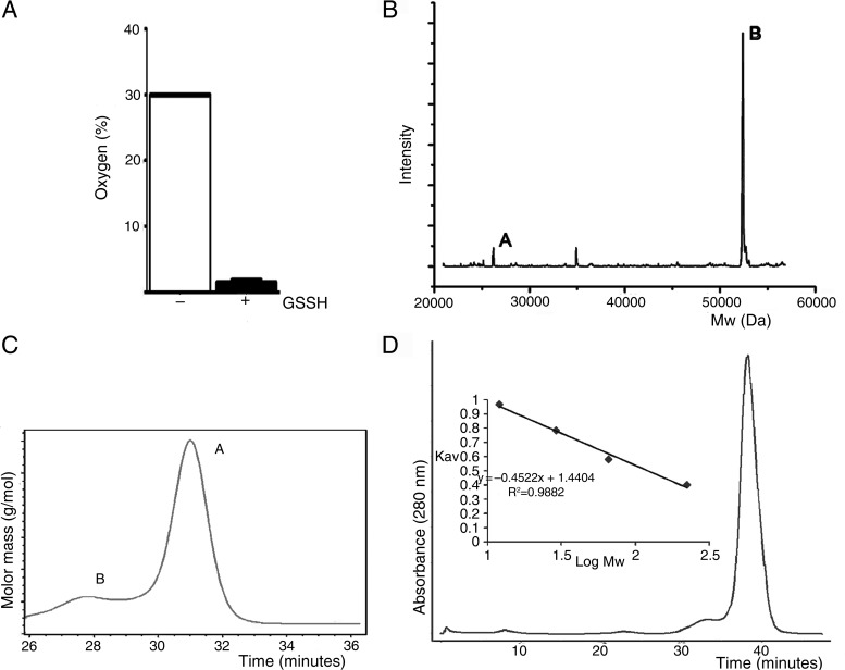
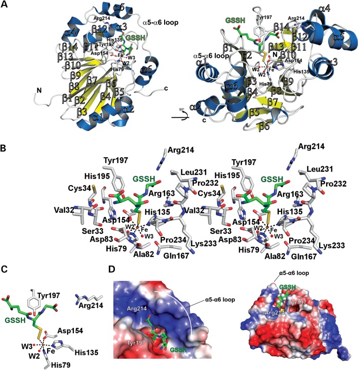
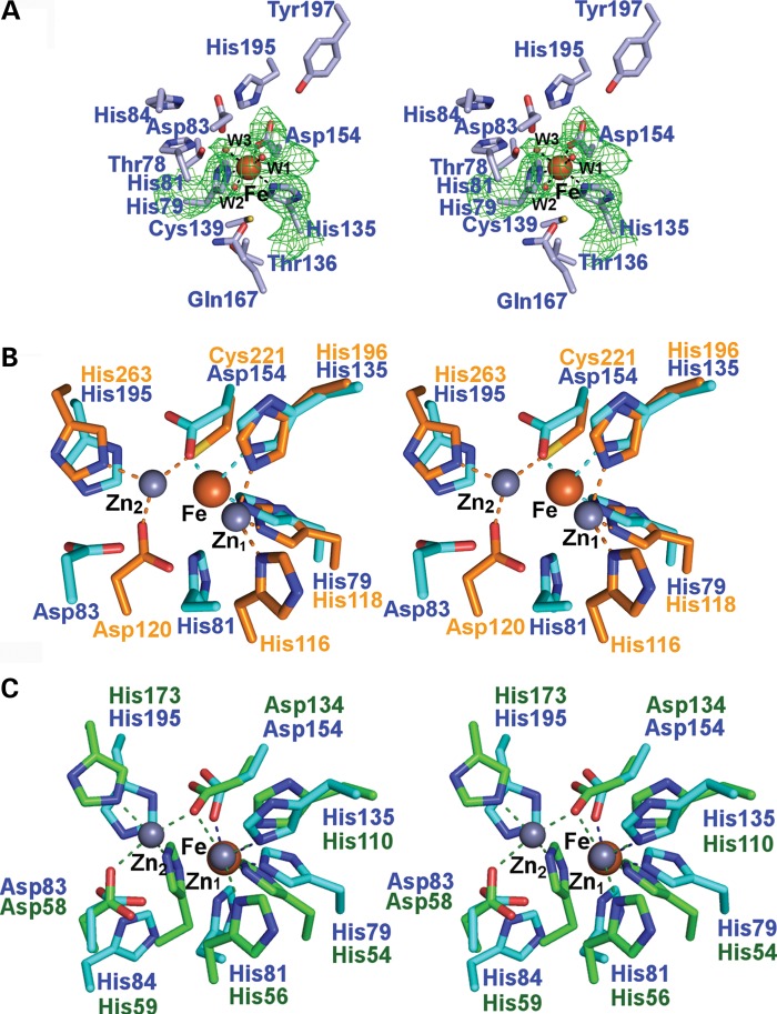
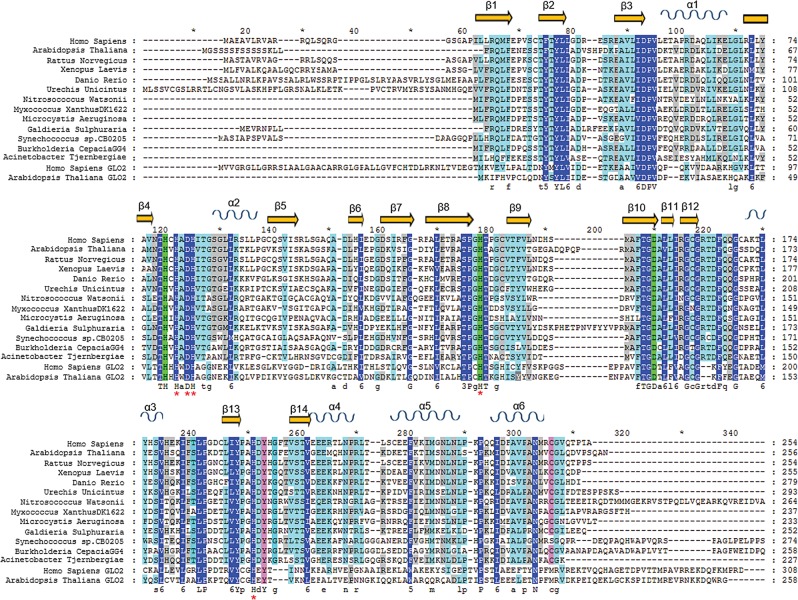
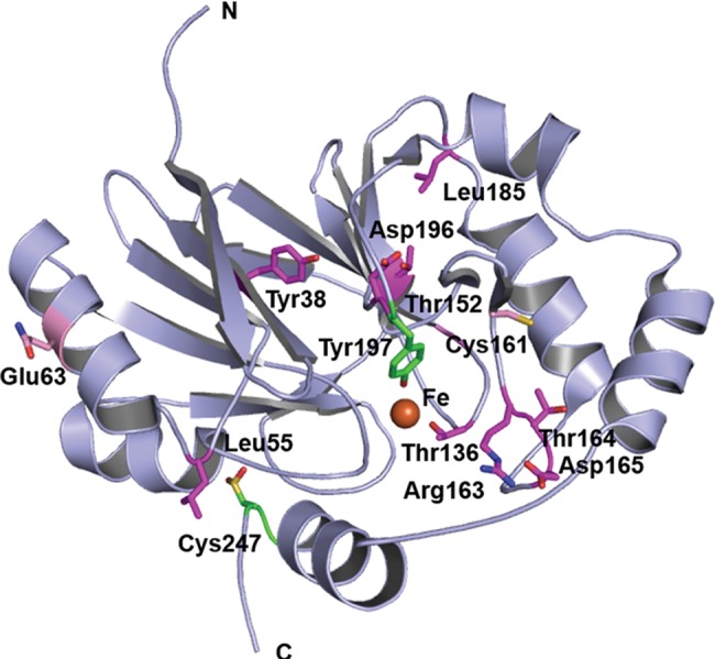
The ethylmalonic encephalopathy protein 1 (ETHE1) catalyses the oxygen-dependent oxidation of glutathione persulfide (GSSH) to give persulfite and glutathione. Mutations to the hETHE1 gene compromise sulfide metabolism leading to the genetic disease ethylmalonic encephalopathy. hETHE1 is a mono-iron binding member of the metallo-β-lactamase (MBL) fold superfamily. We report crystallographic analysis of hETHE1 in complex with iron to 2.6 Å resolution. hETHE1 contains an αββα MBL-fold, which supports metal-binding by the side chains of an aspartate and two histidine residues; three water molecules complete octahedral coordination of the iron. The iron binding hETHE1 enzyme is related to the 'classical' di-zinc binding MBL hydrolases involved in antibiotic resistance, but has distinctive features. The histidine and aspartate residues involved in iron-binding in ETHE1, occupy similar positions to those observed across both the zinc 1 and zinc 2 binding sites in classical MBLs. The active site of hETHE1 is very similar to an ETHE1-like enzyme from Arabidopsis thaliana (60% sequence identity). A channel leading to the active site is sufficiently large to accommodate a GSSH substrate. Some of the observed hETHE1 clinical mutations cluster in the active site region. The structure will serve as a basis for detailed functional and mechanistic studies on ETHE1 and will be useful in the development of selective MBL inhibitors.


Figures

Figure 1.
Figure 1.
Comparison of hETHE1 active site with those of the Class B1, B2 and B3 prokaryotic MBLs. (A) Wall-eyed stereoviews of superimposed active site residues from the Class B1 MBL BcII from Bacillus cereus (PDB ID: 1BVT) (orange), the Class B2 MBL CphA from Aeromonas hydrophila (PDB ID: 3F9O) (pink) and the Class B3 MBL FEZ-1 from Legionella gormanii (PDB ID: 1K07) (blue). The standard BBL numbering system for MBLs is used (17). Residues present in all the three active sites are numbered in black, zinc ions are in light-orange (BcII), light-pink (CphA) and light-blue (FEZ-1). Note that the zinc-ligating residue His121 is only present in the Class B3 FEZ-1, whereas Cys221 is absent in the FEZ-1 B3 MBL compared with the Class B1 and B2 MBLs. The FEZ-1 active site residue composition is most similar to that of the hMBLs, despite the latter apparently displaying closer similarity with the Class B1 MBLs from an overall structural perspective (see Fig. 6). (B) Wall-eyed stereoview of the hETHE1 active site residues. The hETHE1 residue numbering is in blue and based on the enzyme sequence; BBL numbering is shown below in black. Note that in superimposition of hETHE1 with BcII (Fig. 6C), His79ETHE1 (His116BBL) does not correlate with His116BBL of BcII, but with His118BBL showing a different organization of conserved residues in their active sites. Note that the side chains of His84ETHE1 (His121BBL) and FEZ-1 His121BBL are observed in different orientations in their respective active sites, probably because His121BBL of FEZ-1 is involved in an additional metal binding (zinc 2 site), which is not observed in hETHE1.
Figure 2.
Figure 2.
Views from the hETHE1 crystal structure. (A) Wall-eyed stereoview of hETHE1 showing secondary structure elements and the mono iron containing active site. Helices are blue, β-strands yellow and the iron is an orange sphere. (B) Wall-eyed stereoview of the superposition of hETHE1 (blue) and the A. thaliana ETHE1-like enzyme (red) (RMSD 1.43 Å over 230 Cα atoms). The structures reveal very similar overall folds except for small differences in the β2–β3 and β9–β10 loops. (C) The crystallographically observed hETHE1 dimer. Active sites for both chains exist on the same face of the dimer. Chains A and B are in cyan and green, respectively.
Figure 3.
Figure 3.
hETHE1 activity assay and oligomerization state. (A) Oxygen consumption activity assay. hETHE1 activity was measured as percentage of oxygen consumed in the presence of GSSH. Each sample was performed in triplicate. (B) Non-denaturing electrospray soft-ionization mass spectrometry deconvoluted spectrum of purified hETHE1 protein indicates that hETHE1 is primarily dimeric. Peak A (26 170 Da) represents monomer; peak B (52370 Da) the dimer. In both monomeric and dimeric states one iron ion (+56 Da) is bound to each protomer while only in the dimer a +30 Da was observed possibly due to the oxidation state of Cys247 (see the main text and Supplementary Material, Fig. S5) (conditions: 15 µm of hETHE1 in 15 mm ammonium acetate buffer (pH 7.5); cone voltage for the acquisition of the spectra was 80 V). (C) MALS analysis of hETHE1 protein after purification. Peak A (∼51 560 Da) represents monomer; peak B (∼99 190 Da) the tetramer. MALS experiments were carried out by the Biophysical Services of the Biochemistry Department of Oxford University. (D) The molecular mass of hETHE1 in solution was estimated using a Sephadex G250 gel filtration column calibrated with protein standards [beta-amylase (223 kDa), albumin (66 kDa), carbonic anhydrase (29 kDa) and cytochrome C (12 kDa)].
Figure 4.
Figure 4.
hETHE1 surface analysis. (A) Surface representation of the crystallographically observed hETHE1 dimer (chain A cyan, chain B green). Metal binding residues shown as sticks, iron ion shown as orange sphere. (B) Surface representation of the active site groove showing the side chain of Tyr197 directed towards the metal (6.8 Å). (C) The side chain of Cys247 was refined as a sulfinic acid (CSD247) (i.e. RSO2H) as observed in the experimental electron density (3.0 σ mFo-DFc OMIT, omitting oxygen atoms; green mesh). The two serine residues (Ser 88 and Ser 100) are only weakly conserved whilst Cys247 is conserved in most predicted ETHE enzymes as shown in Figure 7.
Figure 5.
Figure 5.
Docking of GSSH in hETHE1 active site. GSSH was manually docked into the active site groove using the shape of the groove and an electrostatic surface potential map as a guide. The GSSH thiol was restrained near the metal ion and the glycyl carboxylate of GSSH was orientated towards the positively charged end of the pocket so that the primary amine on the opposite end of the chain faced the negatively charged surface. The ligand is surrounded by the α5–α6 loop on one side of the groove and by Tyr197 on the other side. All manual docking was performed using Pymol. (A) Ribbons representation of GSSH manually docked into the ETHE1 active site. (B) Wall-eyed stereoview of the hETHE1 active site and surrounding residues possibly participating in substrate binding and/or stabilization. (C) Enlarged view of GSSH docked in the hETHE1 active site. (D) Surface representation of GSSH docked in hETHE1 active site groove. Note the possible substrate interaction with Tyr197 and Arg214.
Figure 6.
Figure 6.
hETHE1 active site and comparison of the hETHE1 crystal structure with other MBL-fold enzymes. (A) Wall-eyed stereoview of the iron binding and active site residues of hETHE1 with representative electron density (3.0 σ mFo-DFc OMIT; green mesh) for side chains of His79 (Nϵ2 to Fe: 2.3 Å), His135 (Nϵ2 to Fe: 2.22 Å), Asp 154 (Oδ2 to Fe: 2.05 Å) and the three water molecules (red spheres) which coordinate (black dashed lines) to the iron (orange sphere). (B) Wall-eyed stereoview of superimposed active site residues from hETHE1 (cyan) and BcII from Bacillus cereus (PDB ID: 1BVT) (orange). The zinc and iron ions are in grey and dark red, respectively. The zinc ions in the Zn1 and Zn2 sites are labelled (17). There is relatively strong conservation in iron-binding residues by ETHE1 and at the Zn1 site of glyoxalase II; although the Zn2 binding site residues are conserved in hETHE1, they do not bind the iron ion. (C) Wall-eyed stereoview of the superimposed active site residues from hETHE1 (cyan) and human glyoxalase II (PDB ID: 1QH3/5) (green). Note. There are more differences between hETHE1 and BcII than between hETHE1 and glyoxalase II.
Figure 7.
Figure 7.
Multiple sequence alignment of ETHE1 from different organisms. COBALT BLAST (36) was used to align sequences identified in a BLAST search (37). The hETHE1 sequence was used as the query and the secondary structure elements are derived from the human ETHE1 structure (PDB ID 4CHL). Secondary structure elements were inserted using the ESPRIT 3 tool (http://espript.ibcp.fr) (38). β-Sheets are shown as yellow arrows, and α-helices as blue sinusoidal waves. Residues are coloured based on conservation: dark blue represents the highest conservation grade, light blue the second highest, grey the third highest and no colour the least conserved. The MBL-fold proteins glyoxalase II from Homo sapiens and Arabidopsis thaliana were added to the multiple sequence alignment. The three iron-binding residues (His79, His135 and Asp154) are highlighted in green; Tyr197 and Cys247 are in pink. Red asterisk (*) indicates a site 2 Zn binding residue of Glyoxalase II (note that Asp134 binds both zinc ions in the Glyoxalase II active site).
Figure 8.
Figure 8.
Clinically observed mutations in hETHE1. Clinically observed substitutions mapped onto the hETHE1 crystal structure. Position of hETHE1 substituted residues (sticks) mapped onto the crystal structure (PDB ID: CHL4). Structure-based sequence analysis using ConSurf reveals that substitutions occur at medium (pink) or highly conserved (magenta) residues (47,48). Tyr197 and the oxidized Cys247 are in green.

Similar articles

Cited by

References

    1. Grosso S., Mostardini R., Farnetani M.A., Molinelli M., Berardi R., Dionisi-Vici C., Rizzo C., Morgese G., Balestri P. (2002) Ethylmalonic encephalopathy: further clinical and neuroradiological characterization. J. Neurol., 249, 1446–1450. - PubMed
    1. Pigeon N., Campeau P.M., Cyr D., Lemieux B., Clarke J.T. (2009) Clinical heterogeneity in ethylmalonic encephalopathy. J. Child. Neurol., 24, 991–996. - PubMed
    1. Barth M., Ottolenghi C., Hubert L., Chrétien D., Serre V., Gobin S., Romano S., Vassault A., Sefiani A., Ricquier D., et al. (2010) Multiple sources of metabolic disturbance in ETHE1-related ethylmalonic encephalopathy. J. Inherit. Metab. Dis., 33, S443–S453. - PubMed
    1. Tiranti V., Briem E., Lamantea E., Mineri R., Papaleo E., De Gioia L., Forlani F., Rinaldo P., Dickson P., Abu-Libdeh B., et al. (2005) ETHE1 mutations are specific to ethylmalonic encephalopathy. J. Med. Genet., 43, 340–346. - PMC - PubMed
    1. Tiranti V., D'Adamo P., Briem E., Ferrari G., Mineri R., Lamantea E., Mandel H., Balestri P., Garcia-Silva M.T., Vollmer B., et al. (2004) Ethylmalonic encephalopathy is caused by mutations in ETHE1, a gene encoding a mitochondrial matrix protein. Am. J. Hum. Genet., 74, 239–252. - PMC - PubMed

Publication types

MeSH terms

Supplementary concepts