. 2015 Mar 15;75(6):1068-79.
doi: 10.1158/0008-5472.CAN-14-2425. Epub 2015 Jan 16.

Tumorigenic activity of merkel cell polyomavirus T antigens expressed in the stratified epithelium of mice

Affiliations

Tumorigenic activity of merkel cell polyomavirus T antigens expressed in the stratified epithelium of mice

Megan E Spurgeon et al. Cancer Res. .

Abstract

Merkel cell polyomavirus (MCPyV) is frequently associated with Merkel cell carcinoma (MCC), a highly aggressive neuroendocrine skin cancer. Most MCC tumors contain integrated copies of the viral genome with persistent expression of the MCPyV large T (LT) and small T (ST) antigen. MCPyV isolated from MCC typically contains wild-type ST but truncated forms of LT that retain the N-terminus but delete the C-terminus and render LT incapable of supporting virus replication. To determine the oncogenic activity of MCC tumor-derived T antigens in vivo, a conditional, tissue-specific mouse model was developed. Keratin 14-mediated Cre recombinase expression induced expression of MCPyV T antigens in stratified squamous epithelial cells and Merkel cells of the skin epidermis. Mice expressing MCPyV T antigens developed hyperplasia, hyperkeratosis, and acanthosis of the skin with additional abnormalities in whisker pads, footpads, and eyes. Nearly half of the mice also developed cutaneous papillomas. Evidence for neoplastic progression within stratified epithelia included increased cellular proliferation, unscheduled DNA synthesis, increased E2F-responsive genes levels, disrupted differentiation, and presence of a DNA damage response. These results indicate that MCPyV T antigens are tumorigenic in vivo, consistent with their suspected etiologic role in human cancer.


Conflict of interest statement

Conflict of Interest: The authors disclose no potential conflicts of interest.

Figures

Figure 1
Figure 1
ROSA26-LSL-MCPyV168 targeted knockin design and expression in stratified squamous epithelium. A) MCPyV168 undergoes alternative splicing to yield wild type ST and truncated LT that contains the LXCXE motif. The ROSA26-LSL-MCPyV168 targeting construct recombined with the ROSA26 genomic locus. K14Cre recombinase-mediated excision of the loxP-stop-loxP cassette results in expression of MCPyV168. Arrows indicate name and approximate position of primers used for ES clone screening and mice genotyping (Table 1). SA, splice acceptor; LSL, loxP-stop-loxP; bpA, bovine polyadenylation sequence. B) PCR genotyping of neonates from a single litter on mixed background with results visualized by agarose gel electrophoresis. C) PCR genotyping of 3 week-old pups on the FVB/N genetic background. D) Adult skin and footpads were harvested from the mice shown in part C and analyzed with antibodies specific to MCPyV LT (Ab3), ST (Ab5), and β-actin. The (+) symbol denotes K14Cre-MCPyV168 mice positive for Cre recombinase and the (−) symbol denotes Cre-negative ROSA26-LSL-MCPyV168 control mice.
Figure 2
Figure 2
Gross epithelial phenotypes of K14Cre-MCPyV168 mice. A) Phenotypes observed on the mixed genetic background. Panel 1) Skin phenotypes are shown on a Cre-negative ROSA26-LSL-MCPyV168 mouse on the left and on a Cre-positive K14Cre-MCPyV168 littermate on the right on postnatal day 8. Panel 2) Skin phenotypes present on a K14Cre-MCPyV168 mouse at postnatal day 18. Panel 3) Representative images of blisters on the footpads of 18 day-old K14Cre-MCPyV168 mice on the mixed genetic background are shown. Panel 4) Shown are enlarged images of representative footpad blisters on K14Cre-MCPyV168 mice. B) Phenotypes observed on the FVB/N genetic background. Panel 1) Skin phenotypes on a Cre-negative ROSA26-LSL-MCPyV168 mouse on the left and on a Cre-positive K14Cre-MCPyV168 littermate on the right on postnatal day 8 are shown. Panel 2) Skin phenotypes observed on a K14Cre-MCPyV168 at postnatal day 18. Whiskers of a ROSA26-LSL-MCPyV168 adult mouse (Panel 3) compared to the whiskers of a K14Cre-MCPyV168 adult mouse (Panel 4). Representative images of eye phenotypes observed on K14Cre-MCPyV168 mice include small eye size (Panel 5) and cataract development (Panel 6). Two representative papillomas are shown on the skin of K14Cre-MCPyV168 mice and indicated by arrowheads in Panel 5 and Panel 6.
Figure 3
Figure 3
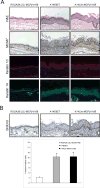
Histological and biomarker analysis of the K14Cre-MCPyV168 stratified epithelium. A) Stratified epithelium of the ear harvested from ROSA26-LSL-MCPyV168 (left), K14E6E7 (middle), and K14Cre-MCPyV168 (right) mice. Tissue was subjected to hematoxylin and eosin (H&E) staining, immunohistochemical analysis for MCM7, and immunofluorescent staining for Keratin 10 (AF594; red) and Keratin 14 (AF594; pseudocolored green). DAPI was used to counterstain cellular nuclei. B) Representative images of BrdU immunohistochemistry on ear epithelium from ROSA26-LSL-MCPyV168 (left), K14E6E7 (middle), and K14Cre-MCPyV168 (right) mice. Quantification of the total percentage of BrdU-positive cells is shown in the bar graph. The total percentage of BrdU-positive cells was calculated using representative samples from groups of ROSA26-LSL-MCPyV168 (n=3), K14E6E7 (n=5), and K14Cre-MCPyV168 (n=5) mice. Error bars = Standard Deviation. *p-value < 0.05 using a two-sided Wilcoxon Rank Sum Test. A black line is used to highlight the basement membrane. All scale bars = 100 μM.
Figure 4
Figure 4
K14Cre-MCPyV168 mice on FVB/N background spontaneously develop papillomas. A) Incidence of wart formation on K14Cre-MCPyV168 mice. As a result of breeding ROSA26-LSL-MCPyV168 and K14Cre mice, 50 out of 98 total mice were positive for Cre recombinase (K14Cre-MCPyV168; red segment of pie chart). Of these K14Cre-MCPyV168 mice, 15/50 mice died prior to weaning (30%; blue portion of bar graph). Of the surviving K14Cre-MCPyV168 mice, 16/35 spontaneously developed papillomas (45.7%; black portion of bar graph). B) Representative image of papillomas arising on the flank and skin surrounding the base of the tail on a K14Cre-MCPyV168 mouse indicated by arrowheads. C) Immunoblot analysis of whole tissue protein lysates isolated from the skin of two independent ROSA26-LSL-MCPyV168 mice and autologous skin and papilloma samples from two independent K14Cre-MCPyV168 mice. Total protein levels were detected using antibodies to MCPyV LT, MCPyV ST, and β-actin as a loading control. The (+) symbol denotes K14Cre-MCPyV168 mice positive for Cre recombinase and the (−) symbol denotes Cre-negative ROSA26-LSL-MCPyV168 control mice. D) Histological and biomarker analysis of a papilloma harvested from the skin of a K14Cre-MCPyV168 mouse and age/location-matched tissue from a control ROSA26-LSL-MCPyV168 mouse. Representative images of H&E, co-immunofluorescence analysis of keratin 10 (AF594; red) and keratin 14 (AF488; green), and MCM7 immunohistochemistry are shown. DAPI was used to counterstain cellular nuclei. Scale bars = 100 μM.
Figure 5
Figure 5
Interrogation of molecular pathways in K14Cre-MCPyV168 skin and papillomas. A) Immunoblot analysis of whole tissue lysates isolated from the skin of two independent ROSA26-LSL-MCPyV168 mice and autologous skin samples and papillomas of two independent K14Cre-MCPyV168 mice using antibodies to MCPyV LT, survivin, and β-actin as a loading control. B) Representative images of hematoxylin and eosin (H&E) and immunofluorescence staining on tissue sections from age and location-matched ROSA26-LSL-MCPyV168 skin (left) and either non-papilloma associated skin (middle) or a papilloma (right) harvested from a K14Cre-MCPyV168 mouse. Immunofluorescence analysis for keratin 14 (AF488; green), keratin 10 (AF594; red), and γ-H2AX staining (AF488; green) were performed on immediately adjacent tissue sections, while pS1981-ATM staining (pATM) (AF488; pseudocolored red) was performed on a non-adjacent tissue section. DAPI was used to counterstain cellular nuclei. The (+) symbol denotes mice positive for Cre recombinase and the (−) symbol denotes Cre-negative mice. Scale bars = 100 μM.
Figure 6
Figure 6
Merkel cells are positive for MCM7 in K14Cre-MCPyV168 mice. A) Immunofluorescence analysis showing co-localization of cytokeratin 8 (CK8) and cytokeratin 20 (CK20) in Merkel cells. Tissue sections of whisker pads harvested from K14Cre-MCPyV168 adult mice were analyzed by co-immunofluorescence staining for CK8 (AF647; pink) and CK20 (AF488; green). DAPI was used to counterstain cellular nuclei. Microscopic images were taken of the whisker vibrissae to highlight Merkel cells along the vibrissae follicle. B) Sequential sections of whisker pad tissue harvested from ROSA26-LSL-MCPyV168 and K14Cre-MCPyV168 adult mice were analyzed by immunofluorescence for CK8 (AF647; pink) and MCM7 (AF488; green). Scale bars in 20X images = 100 μM, scale bars in 63X images = 10 μM.

Similar articles

Cited by

References

    1. Feng H, Shuda M, Chang Y, Moore PS. Clonal integration of a polyomavirus in human Merkel cell carcinoma. Science. 2008;319(5866):1096–100. - PMC - PubMed
    1. DeCaprio JA, Garcea RL. A cornucopia of human polyomaviruses. Nature reviews Microbiology. 2013;11(4):264–76. - PMC - PubMed
    1. Carter JJ, Daugherty MD, Qi X, Bheda-Malge A, Wipf GC, Robinson K, et al. Identification of an overprinting gene in Merkel cell polyomavirus provides evolutionary insight into the birth of viral genes. Proc Natl Acad Sci U S A. 2013;110(31):12744–9. - PMC - PubMed
    1. Shuda M, Feng H, Kwun HJ, Rosen ST, Gjoerup O, Moore PS, et al. T antigen mutations are a human tumor-specific signature for Merkel cell polyomavirus. Proc Natl Acad Sci U S A. 2008;105(42):16272–7. - PMC - PubMed
    1. Tolstov YL, Knauer A, Chen JG, Kensler TW, Kingsley LA, Moore PS, et al. Asymptomatic primary Merkel cell polyomavirus infection among adults. Emerg Infect Dis. 2011;17(8):1371–80. - PMC - PubMed

Publication types