. 2015 Mar 15;75(6):1035-45.
doi: 10.1158/0008-5472.CAN-13-1625. Epub 2015 Jan 16.

ERBB3-independent activation of the PI3K pathway in EGFR-mutant lung adenocarcinomas

Affiliations

ERBB3-independent activation of the PI3K pathway in EGFR-mutant lung adenocarcinomas

Xiaoling Song et al. Cancer Res. .

Abstract

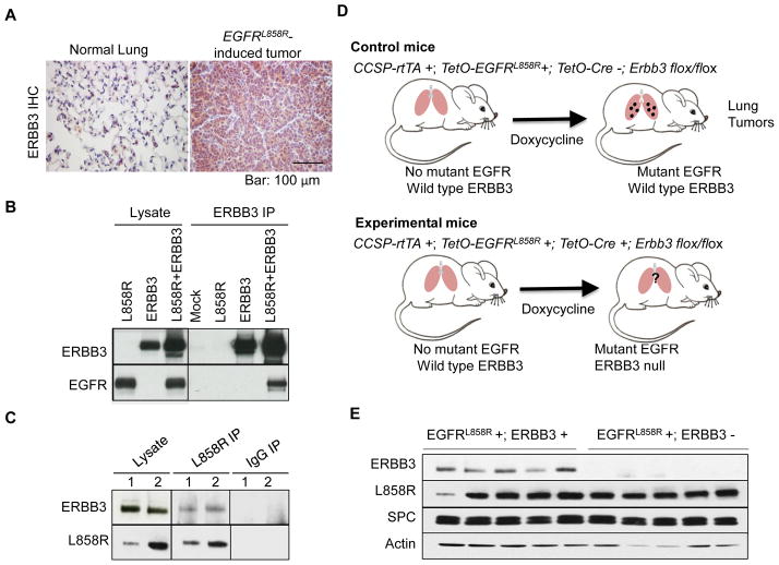
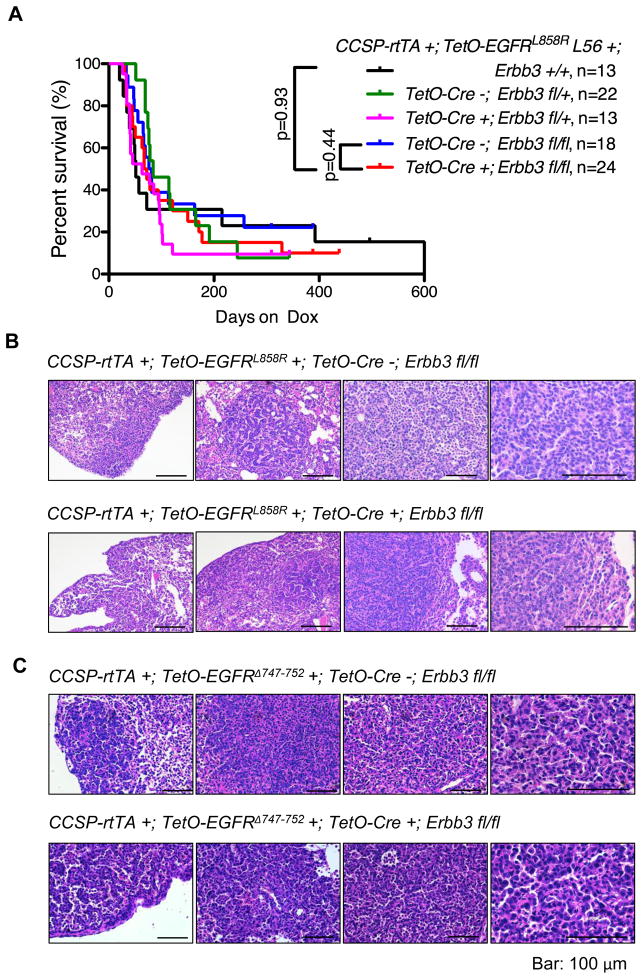
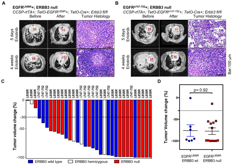
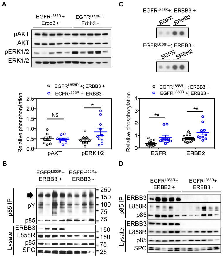
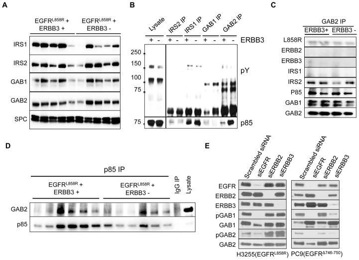
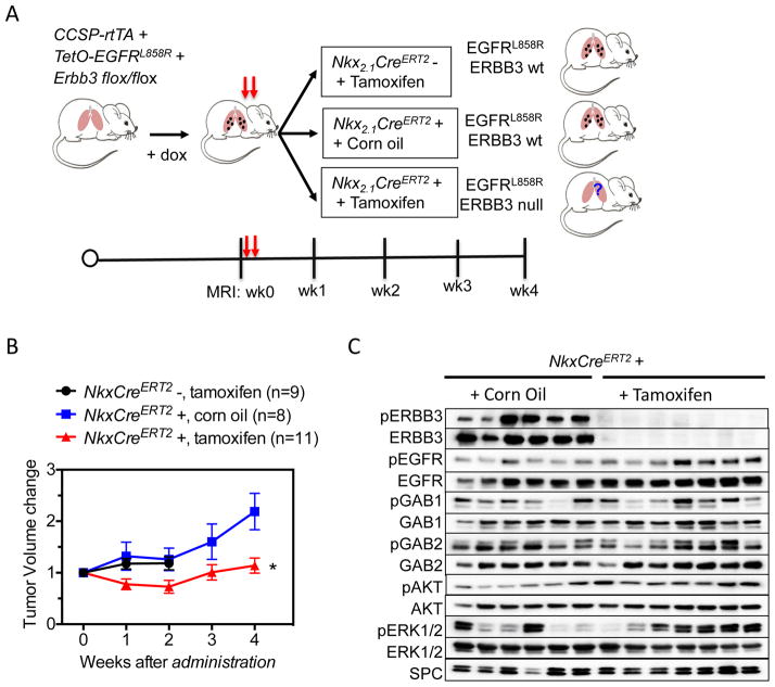
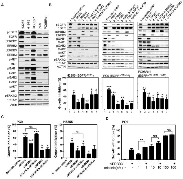
ERBB3, a member of the EGFR family of receptor tyrosine kinases, has been implicated in activation of the PI3K pathway in human lung adenocarcinomas driven by EGFR mutations. We investigated the contribution of ERBB3 to the initiation, progression, and therapeutic response of EGFR-induced lung adenocarcinomas using tetracycline- and tamoxifen-inducible transgenic mouse models. Deletion of Erbb3 at the time of induction of mutant EGFR had no effect on tumorigenesis, demonstrating that ERBB3 is not required to initiate tumorigenesis. Tumors that developed in the absence of ERBB3 remained sensitive to EGFR tyrosine kinase inhibitors and retained activation of the PI3K-AKT pathway. Interestingly, acute loss of Erbb3 suppressed further growth of established EGFR(L858R)-mediated lung tumors. Four weeks after deletion of Erbb3, the tumors exhibited phosphorylation of EGFR, of the adaptor proteins GAB1 and GAB2, and of the downstream signaling molecules AKT and ERK, suggesting that alternative signaling pathways could compensate for loss of Erbb3. Similar to our observations with mouse tumors, we found that GAB adaptor proteins play a role in ERBB3-independent activation of the PI3K pathway by mutant EGFR in EGFR-mutant human cell lines. Finally, in such cell lines, increased levels of phosphorylation of ERBB2 or MET were associated with reduced sensitivity to acute loss of ERBB3, suggesting remarkable plasticity in the signaling pathways regulated by mutant EGFR with important therapeutic implications.


Conflict of interest statement

Conflicts of Interest: Rights to a patent application for EGFRT790M testing were licensed on behalf of KP and HV by Memorial Sloan-Kettering Cancer Center to MolecularMD. KP has served as a consultant for Takeda and her lab receives research support from AstraZeneca.

Figures

Figure 1
Figure 1. ERBB3 is present in murine EGFR mutant lung adenocarcinomas and interacts with mutant EGFR
(A) Immunohistochemical staining for ERBB3 is strong in EGFRL858R-induced lung tumors (right) but weak or non-existent in normal lung from control mice (left). (B) EGFRL858R interacts with ERBB3 in transfected cells. Plasmids encoding human EGFRL858R, mouse ERBB3 or both were transfected into 293T cells. Two days after transfection, an antibody to ERBB3 was used to co-precipitate ERBB3 and proteins associated with it. Immunoprecipitates were blotted with antibodies to EGFR and ERBB3. Results from mock-transfected cells are shown as indicated. (C) Mutant EGFRL858R interacts with ERBB3 in mutant EGFR-induced lung tumors. EGFRL858R or IgG immunoprecipitates from two CCSP-rtTA+;TetO-EGFRL858R+ mice (1 and 2) were immunoblotted with antibodies specific to mutant EGFRL858R and ERBB3. (D) Strategy used to conditionally delete Erbb3 in type II pneumocytes in tetracycline-inducible mouse models of EGFR mutant lung cancer. Control mice (top) express mutant EGFR and wild-type Erbb3 in the lung epithelium upon induction of doxycycline. Experimental mice (bottom) express mutant EGFR but not ERBB3 due to the presence of Cre recombinase, which deletes floxed sequences in the Erbb3 locus. (E) ERBB3 protein is not detected in lungs of mice upon deletion of Erbb3. Immunoblots of lung protein extracts with antibodies to EGFRL858R, ERBB3, Surfactant Protein C (SPC) and Actin are shown. Protein levels were normalized for numbers of pneumocytes as judged by levels of surfactant protein C. EGFRL858R+; Erbb3+ (control mice) and EGFRL858R+; Erbb3− (experimental mice). IP, immunoprecipitation.
Figure 2
Figure 2. Development of lung adenocarcinomas in mice lacking ERBB3
(A) Kaplan-Meier survival curves comparing CCSP-rtTA+;TetO-EGFRL858R+ mice with (Erbb3 +/+, Erbb3fl/fl; Cre−, Erbb3 fl/+; Cre−), without (Erbb3 fl/fl; Cre+) or heterozygous for (Erbb3 fl/+; Cre+) Erbb3. L56, transgenic line 56. (B) Hematoxylin and Eosin staining of EGFRL858R-induced tumors that developed in the presence and absence of ERBB3. Representative images of mouse lungs from mice on doxycycline for approximately two months are shown. Bar, 100μm. (C) Hematoxylin and Eosin staining of EGFRΔ747-752-induced tumors that developed in the presence and absence of ERBB3. Representative images of mouse lungs from mice on doxycycline for approximately four months are shown. Bar, 100μm.
Figure 3
Figure 3. Lung tumors that develop in the absence of ERBB3 are sensitive to erlotinib
(A) CCSP-rtTA +; TetO-EGFRL858R+; TetO-Cre +; Erbb3fl/fl mice respond to short- and long-term erlotinib treatment. Sick mice were treated with erlotinib for 5 days or 4 weeks before they were sacrificed. Mice were imaged prior to (before) and towards the end (after) of treatment. Representative MR images and H&E stained sections are shown. Images of residual tumors observed by histopathology are shown. Bar, 100μm. (B) CCSP-rtTA +; TetO-EGFRΔ747-752+; TetO-Cre +; Erbb3fl/fl mice respond to short- and long-term erlotinib treatment. Sick mice were treated with erlotinib for 5 days or 4 weeks before they were sacrificed. Mice were imaged prior to (before) and towards the end (after) of treatment. Representative MR images and H&E stained sections are shown. Images of residual tumors observed by histopathology are shown. Bar, 100μm. (C) Waterfall plot showing the change in tumor volume from baseline in mice with mutant EGFR-induced lung tumors (EGFRL858R or EGFRΔ747-752) upon treatment with erlotinib for 4 weeks. The Erbb3 genotypes of individual mice are indicated. (D) Lung tumor volume change in mice with EGFRL858R-induced lung tumors upon treatment with erlotinib for 4 weeks.
Figure 4
Figure 4. Intact PI3K and MAPK Signaling in Lung Tumors Deficient for ERBB3
(A) Immunoblots of protein lysates from lungs of mice with EGFRL858R-induced lung tumors arising in the presence (EGFRL858R+; ERBB3+) or absence (EGFRL858R+; ERBB3−) of ERBB3 and probed with antibodies to pAKT, total AKT, pERK and total ERK (top). The data are presented as the mean ± the standard error (bottom). (B) Immunoprecipitation of p85 from EGFRL858R-induced lung tumors. The immunoprecipitates were blotted with anti-phosphotyrosine and p85 antibodies. The arrow indicates bands the size of EGFR family receptors in the phosphotyrosine immunoblot. Immunoblots of lysates using antibodies to ERBB3, EGFR, p85 and SPC are shown. (C) Phospho-RTK arrays reveal increased phosphorylation of EGFR and ERBB2 upon deletion of ERBB3. Whole lung tumor lysates from eight ERBB3 wild type mice and ten ERBB3 null mice were hybridized to phospho-RTK arrays (top). Signals for EGFR family members on representative arrays are shown. Data are presented as the mean ± standard error (bottom). (D) Immunoprecipitation of p85 from lung tumor lysates that develop in the presence (EGFRL858R+; ERBB3+) and absence (EGFRL858R+; ERBB3−) of Erbb3. Immunoprecipitates were immunoblotted with antibodies to ERBB3, EGFRL858R and p85. IP, immunoprecipitation. *indicates p-value ≤0.05; ** indicates p-value < 0.01; NS, not significant.
Figure 5
Figure 5. Adapter proteins bind p85 in vivo in EGFR mutant lung tumors
(A) Immunoblots of lung protein lysates from mice with EGFRL858R-induced tumors arising in the presence (EGFRL858R+; ERBB3+) and absence (EGFRL858R+; ERBB3−) of ERBB3 using antibodies to proteins that can directly bind p85 (IRS1, IRS2, GAB1 and GAB2). The same lysates were blotted with an antibody to SPC as a control for the fraction of pneumocytes present. (B) Co-immunoprecipitation of the adapter proteins IRS1, IRS2, GAB1 and GAB2 from lung protein lysates from mice with EGFRL858R-induced tumors arising in the presence (+) and absence (−) of ERBB3. The immunoprecipitates were blotted with antibodies to phosphotyrosine (pY, top) and p85 (bottom). (C) Co-immunoprecipitation of EGFR, GAB1, GAB2, IRS1, IRS2 and p85, with GAB2 from lung protein lysates from mice with EGFRL858R-induced tumors arising in the presence (+) and absence (−) of Erbb3. (D) Co-immunoprecipitation of GAB2 with p85 from lung protein lysates from mice with EGFRL858R-induced tumors arising in the presence (+) and absence (−) of Erbb3. IP, immunoprecipitation. (E) GAB1 and GAB2 are expressed in EGFR mutant lung cancer cell lines and phosphorylated by EGFR. H3255 and PC9 cells were transfected with either scrambled siRNA or siRNA of EGFR, ERBB2 or ERBB3. Total and phospho- protein levels were analyzed by Western blotting.
Figure 6
Figure 6. Partial dependence of EGFRL858R-induced lung adenocarcinomas on Erbb3
(A) Model for tamoxifen induced deletion of Erbb3. Mutant EGFRL858R tetra transgenic mice were fed doxycycline to induce tumors. When the tumor size reached a minimum volume of 100 mm3, experimental mice (Nkx2.1-CreERT2+) were administered tamoxifen by oral gavage at the doses of 250 mg/day for two days. Control animals were Nkx2.1-CreERT2 negative and treated with tamoxifen or Nkx2.1-CreERT2 positive treated with corn oil. After tamoxifen treatment, mice were imaged using MRI weekly for 4 weeks. The tumor burden for each group is shown in Fig. 6B. Mouse lungs were collected at the end of 4 weeks or when moribund and tumor extracts were analyzed by Western blot for the indicated proteins (Fig. 6C).
Figure 7
Figure 7. ERBB3 knockdown partially affects the viability of EGFR mutant, human lung cancer cell lines
(A) Western blots of protein lysates derived from EGFR mutant lung cancer cell lines probed with antibodies as indicated. (B) H3255, PC9 and PC9BRc1 cells were transiently transfected with pools of siRNAs against EGFR, ERBB3, GAB1, GAB1+ERBB3, GAB1+GAB2, GAB1+GAB2+ERBB3 or a scrambled siRNA control as indicated. Three days after transfection, cells were lysed in RIPA buffer and proteins were analyzed by Western blotting and cell viability was examined using the CellTiter blue assay. Experiments were performed at least three times for each cell line. (C) Viability of PC9 and H3255 cells after transfection with pools of siRNAs against EGFR, ERBB3, EGFR+ERBB3, ERBB2 and ERBB2+ERBB3. Cell viability was measured using the Cell Titer Blue Assay. (D) Growth inhibition in PC9 cells upon ERBB3 knockdown treated with varying concentrations of erlotinib as indicated. *indicates p-value ≤ 0.05, *** indicates p-value ≤ 0.01. NS: not significant.

References

    1. Riese DJ, 2nd, Stern DF. Specificity within the EGF family/ErbB receptor family signaling network. BioEssays : news and reviews in molecular, cellular and developmental biology. 1998;20(1):41–8. - PubMed
    1. Guy PM, Platko JV, Cantley LC, Cerione RA, Carraway KL., 3rd Insect cell-expressed p180erbB3 possesses an impaired tyrosine kinase activity. Proceedings of the National Academy of Sciences of the United States of America. 1994;91(17):8132–6. - PMC - PubMed
    1. Lynch TJ, Bell DW, Sordella R, Gurubhagavatula S, Okimoto RA, Brannigan BW, et al. Activating mutations in the epidermal growth factor receptor underlying responsiveness of non-small-cell lung cancer to gefitinib. The New England journal of medicine. 2004;350(21):2129–39. - PubMed
    1. Paez JG, Janne PA, Lee JC, Tracy S, Greulich H, Gabriel S, et al. EGFR mutations in lung cancer: correlation with clinical response to gefitinib therapy. Science (New York, NY. 2004;304(5676):1497–500. - PubMed
    1. Pao W, Miller V, Zakowski M, Doherty J, Politi K, Sarkaria I, et al. EGF receptor gene mutations are common in lung cancers from “never smokers” and are associated with sensitivity of tumors to gefitinib and erlotinib. Proceedings of the National Academy of Sciences of the United States of America. 2004;101(36):13306–11. - PMC - PubMed

Publication types