Plasma cell gingivitis with severe alveolar bone loss
- PMID: 25596294
- PMCID: PMC4307090
- DOI: 10.1136/bcr-2014-207013
Plasma cell gingivitis with severe alveolar bone loss
Abstract
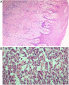
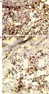
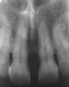
Plasma cell gingivitis is a rare benign condition of the gingiva characterised by sharply demarcated erythaematous and oedematous gingiva often extending up to the muco gingival junction. It is considered a hypersensitive reaction. It presents clinically as a diffuse, erythaematous and papillary lesion of the gingiva, which frequently bleeds, with minimal trauma. This paper presents a case of a 42-year-old man who was diagnosed with plasma cell gingivitis, based on the presence of plasma cells in histological sections, and severe alveolar bone loss at the affected site, which was managed by surgical intervention.
2015 BMJ Publishing Group Ltd.
Figures


Similar articles
-
Plasma cell gingivitis: a case report.JNMA J Nepal Med Assoc. 2012 Apr-Jun;52(186):85-7. JNMA J Nepal Med Assoc. 2012. PMID: 23478737
-
Plasma cell gingivitis: treatment with chlorpheniramine maleate.Int J Periodontics Restorative Dent. 2015 May-Jun;35(3):411-3. doi: 10.11607/prd.2111. Int J Periodontics Restorative Dent. 2015. PMID: 25909529
-
Plasma cell gingivitis related to the use of herbal toothpaste.Br Dent J. 1989 May 20;166(10):375-6. doi: 10.1038/sj.bdj.4806848. Br Dent J. 1989. PMID: 2736170
-
Plasma cell gingivitis: does it exist? Report of a case and review of the literature.SADJ. 2008 Aug;63(7):394-5. SADJ. 2008. PMID: 19054906 Review.
-
Plasma Cell Gingivitis and Its Mimics.Oral Maxillofac Surg Clin North Am. 2023 May;35(2):261-270. doi: 10.1016/j.coms.2022.10.003. Epub 2023 Feb 15. Oral Maxillofac Surg Clin North Am. 2023. PMID: 36805902 Review.
Cited by
-
Immunoglobulin G4-related periodontitis: case report and review of the literature.BMC Oral Health. 2021 May 28;21(1):279. doi: 10.1186/s12903-021-01592-2. BMC Oral Health. 2021. PMID: 34049546 Free PMC article. Review.
-
Clinico-Pathological Profile and Outcomes of 45 Cases of Plasma Cell Gingivitis.J Clin Med. 2021 Feb 18;10(4):830. doi: 10.3390/jcm10040830. J Clin Med. 2021. PMID: 33670562 Free PMC article.
-
Pitfalls and Challenges in Oral Plasma Cell Mucositis: A Systematic Review.J Clin Med. 2022 Nov 4;11(21):6550. doi: 10.3390/jcm11216550. J Clin Med. 2022. PMID: 36362778 Free PMC article. Review.
-
An atypical presentation of plasma cell gingivitis with generalized skin lesions.J Oral Maxillofac Pathol. 2021 Mar;25(Suppl 1):S54-S57. doi: 10.4103/jomfp.JOMFP_292_20. Epub 2021 Mar 19. J Oral Maxillofac Pathol. 2021. PMID: 34083972 Free PMC article.
-
A Rare Case of Plasma Cell Gingivitis with Cheilitis.Case Rep Dent. 2019 Dec 16;2019:2939126. doi: 10.1155/2019/2939126. eCollection 2019. Case Rep Dent. 2019. PMID: 31934461 Free PMC article.
References
-
- Bhaskar SN, Levin MP, Frisch J. Plasma cell granuloma of periodontal tissues. Report of 45 cases. Periodontics 1968;6:272–6. - PubMed
-
- Shafer WG, Hine MK, Levy BM. Shafer's textbook of oral pathology. 5th edn Philladelphia: Saunders Co, 2006:546.
Publication types
MeSH terms
LinkOut - more resources
Full Text Sources
Other Literature Sources
