

Amyloidosis of the renal pelvis: a harbinger of mammary carcinoma?
- PMID: 25596296
- PMCID: PMC4307064
- DOI: 10.1136/bcr-2014-207955
Amyloidosis of the renal pelvis: a harbinger of mammary carcinoma?
Abstract
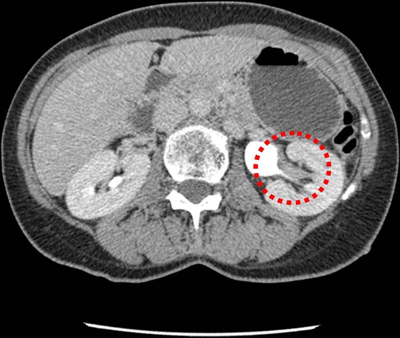
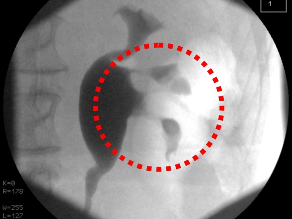
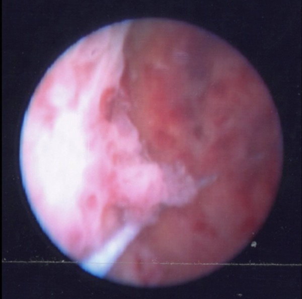
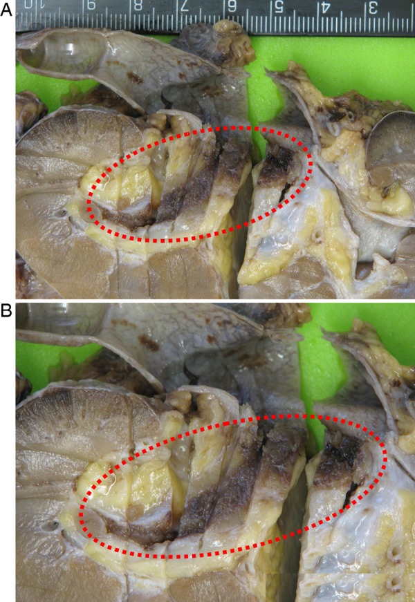
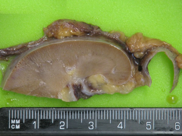
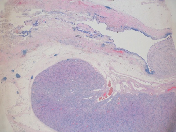
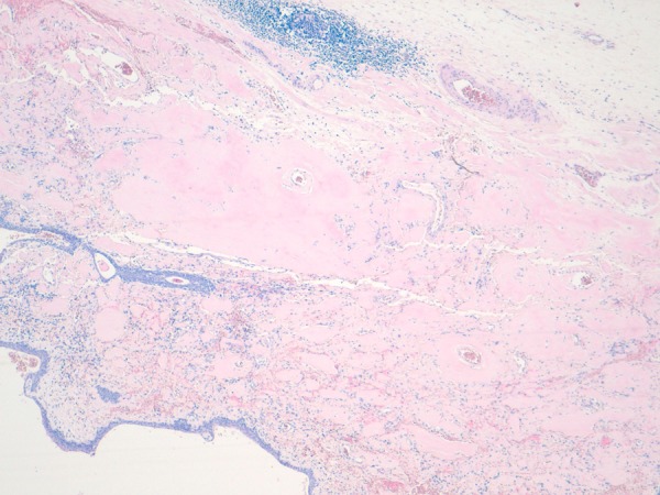
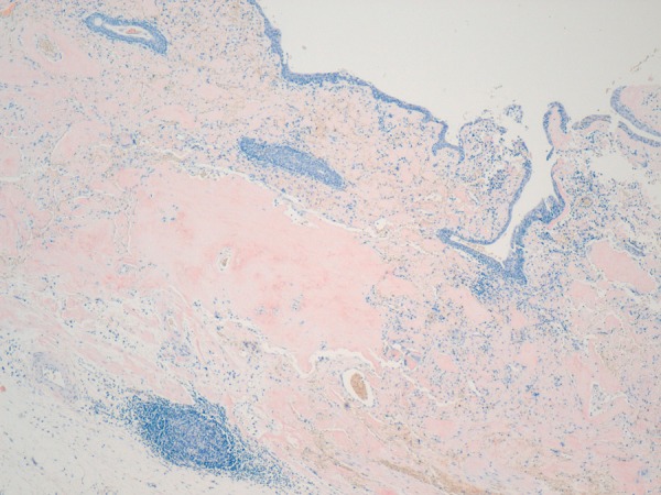
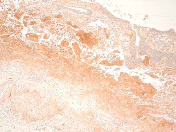
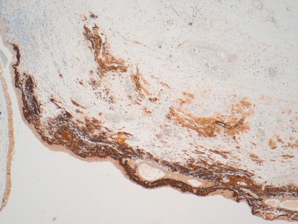
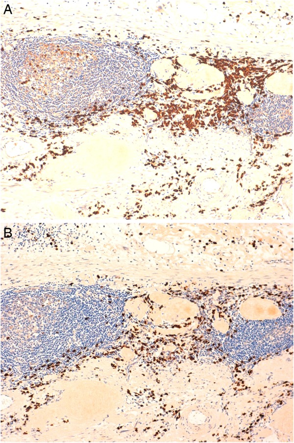
We describe a rare case of light chain immunoglobulin amyloid (AL) accumulation in the central and lower pole renal calyces. Our patient, a woman aged 60, presented with several episodes of gross haematuria. Radiological imaging detected a filling defect in the left renal pelvis. Rigid ureteroscopy showed a corresponding mucosal abnormality resembling transitional cell carcinoma. A definitive preoperative tissue diagnosis could not be reached. Laparoscopic-assisted left nephroureterectomy was indicated. Histopathological examination excluded malignancy, revealing congophilic deposits of submucosal amyloid. A constellation of findings confirmed localised or primary amyloidosis with an AL immunophenotype but no evidence of clonal B-cell disease in the amyloid-associated lymphoplasmacytic cell infiltrate. Investigation for systemic plasma cell dyscrasia and echocardiography and scintigraphy for visceral amyloid deposits were negative for systemic disease. At a follow-up period of 30 months, there is no recurrence. However, our patient was diagnosed with breast cancer 21 months ago.
2015 BMJ Publishing Group Ltd.
Figures
References
-
- Wetzel R. Kinetics and thermodynamics of amyloid fibril assembly. Acc Chem Res 2006;39:671–9. - PubMed
-
- Gertz MA, Comenzo R, Falk RH et al. Definition of organ involvement and treatment response in immunoglobulin light chain amyloidosis (AL): a consensus opinion from the 10th International Symposium on Amyloid and Amyloidosis, Tours, France, 18–22 April 2004. Am J Hematol 2005;79:319–28. - PubMed
-
- Borza T, Shah RB, Faerber GJ et al. Localized amyloidosis of the upper urinary tract: a case series of three patients managed with reconstructive surgery or surveillance. J Endourol 2010;24:641–4. - PubMed
-
- Dias R, Fernandes M, Patel RC et al. Amyloidosis of renal pelvis and urinary bladder. Urology 1979;14:401–4. - PubMed
Publication types
MeSH terms
Substances
LinkOut - more resources
Full Text Sources
Other Literature Sources
Medical