. 2015 Jul 1;212(1):157-65.
doi: 10.1093/infdis/jiv020. Epub 2015 Jan 16.

Interleukin 10-Dominant Immune Response and Increased Risk of Cutaneous Leishmaniasis After Natural Exposure to Lutzomyia intermedia Sand Flies

Affiliations

Interleukin 10-Dominant Immune Response and Increased Risk of Cutaneous Leishmaniasis After Natural Exposure to Lutzomyia intermedia Sand Flies

Augusto M Carvalho et al. J Infect Dis. .

Abstract

Background: Leishmaniasis is caused by parasites transmitted to the vertebrate host by infected sand flies. During transmission, the vertebrate host is also inoculated with sand fly saliva, which exerts powerful immunomodulatory effects on the host's immune response.

Methods: We conducted a prospective cohort analysis to characterize the human immune response to Lutzomyia intermedia saliva in 264 individuals, from an area for cutaneous leishmaniasis (CL) caused by Leishmania braziliensis.

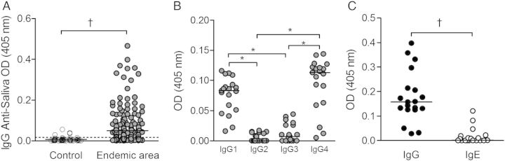
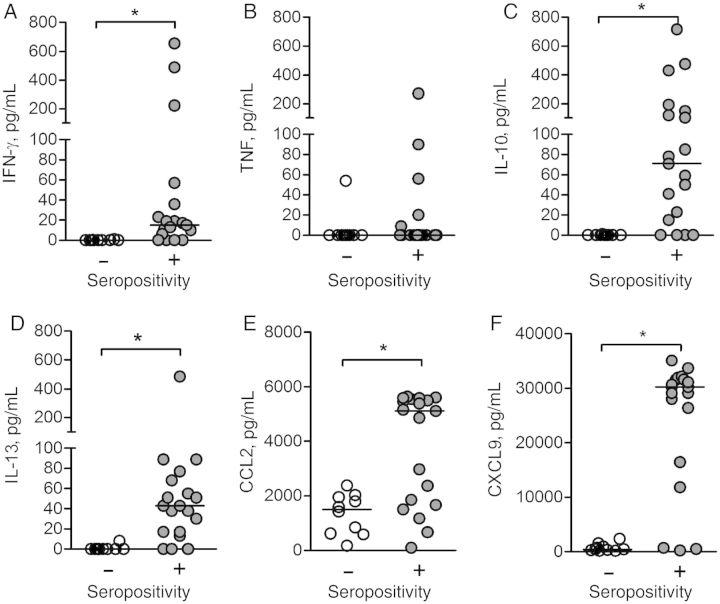
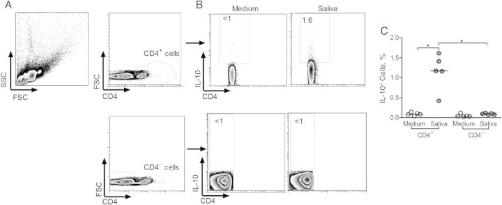
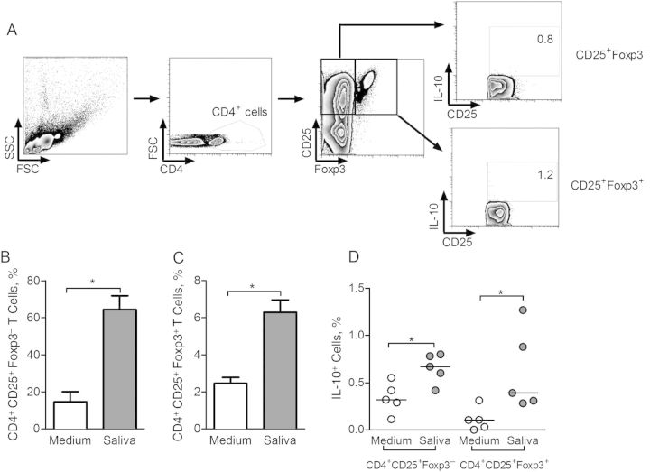
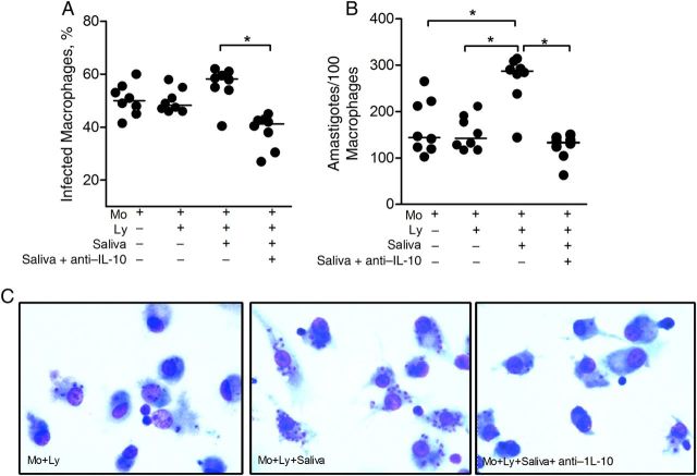
Results: Antibodies were found in 150 individuals (56.8%); immunoglobulin G1 and G4 were the predominant subclasses. Recall responses to salivary gland sonicate showed elevated production of interleukin 10 (IL-10), interleukin 13, interferon γ, CXCL9, and CCL2 compared with controls. CD4(+)CD25(+) T cells, including Foxp3(+) cells, were the main source of IL-10. L. braziliensis replication was increased (P < .05) in macrophages cocultured with saliva-stimulated lymphocytes from exposed individuals and addition of anti-IL-10 reverted this effect. Positive correlation between antibody response to saliva and cellular response to Leishmania was not found. Importantly, individuals seropositive to saliva are 2.1 times more likely to develop CL (relative risk, 2.1; 95% confidence interval, 1.07-4.2; P < .05).

Conclusions: Exposure to L. intermedia sand flies skews the human immune response, facilitating L. braziliensis survival in vitro, and increases the risk of developing CL.

Keywords: ELISA; L. braziliensis; chemokines; cutaneous leishmaniasis; cytokines; killing assay; lutzomyia intermedia; sand fly saliva.


Figures

Figure 1.
Figure 1.
Humoral immune response to Lutzomyia intermedia saliva in a cutaneous leishmaniasis (CL)-endemic area. A, Total immunoglobulin (Ig) G response in residents of a nonendemic area (n = 46) (white circles) and a CL-endemic area (n = 264) (gray circles). B, IgG subclasses in a subset of residents (n = 19) from the endemic area represented in A who were seropositive to L. intermedia saliva. C, Comparison of IgG and IgE levels in the same subset. Circles represent individual values; horizontal lines, median optical density (OD) values; dotted line in A, cutoff level. *P < .05; P < .001.
Figure 2.
Figure 2.
Detection of Lutzomyia intermedia salivary proteins by Western blot analysis. L. intermedia saliva was submitted to sodium dodecyl sulfate polyacrylamide gel electrophoresis and tested against serum samples from residents (n = 5) of a cutaneous leishmaniasis endemic area.
Figure 3.
Figure 3.
Cellular immune response to Lutzomyia intermedia saliva in a cutaneous leishmaniasis-endemic area. Peripheral blood mononuclear cells from a subset of residents who were seronegative (n = 10) (white circles) or seropositive (n = 19) (gray circles) to sand fly saliva were restimulated with L. intermedia saliva (equivalent to 1.5 pair) for 72 hours. Cytokine levels of interferon (IFN) γ (A), tumor necrosis factor (TNF) (B), interleukin 10 (IL-10) (C), interleukin 13 (IL-13) (D), CCL2 (E), and CXCL9 (F) were determined by enzyme-linked immunosorbent assay after 72 hours. Horizontal lines indicate median levels. *P < .05.
Figure 4.
Figure 4.
Frequency of interleukin 10 (IL-10)–producing cells in individuals exposed to Lutzomyia intermedia sand flies in a cutaneous leishmaniasis–endemic area. A, Gating strategy for flow cytometry analysis of CD4+ and CD4 cells after restimulation of peripheral blood mononuclear cells with L. intermedia saliva. In this sample gating, cells were first gated for size and granularity (FSC × SSC). The gated cells were further analyzed for expression of CD4. B, IL-10 expression was determined in both CD4+ and CD4 gated populations after culture in medium alone (control) or in the presence of L. intermedia saliva. C, Frequency of IL-10+ cells in CD4 and in CD4+ populations, in control (white circles) and saliva-stimulated (gray circles) cultures from 5 seropositive individuals. Circles represent individual values; horizontal lines, indicate median levels. *P < .05. Abbreviations: FSC, forward scattered light; SSC, side scattered light.
Figure 5.
Figure 5.
Frequency of CD4+CD25+Foxp3 and CD4+CD25+Foxp3+ T cells in individuals exposed to Lutzomyia intermedia sand flies in a cutaneous leishmaniasis-endemic area. A, Gating strategy for flow cytometry analysis of CD4+CD25+Foxp3+ cells after restimulation with L. intermedia saliva. In this sample gating, cells were first gated for size and granularity (FSC × SSC). CD25 and Foxp3 expression were determined in CD4+ gated cells and, within these, interleukin 10 (IL-10) expression was determined. B, Frequency of CD4+CD25+Foxp3 cells after culture in medium alone (white bar) or in the presence of L. intermedia saliva (gray bar). C, Frequency of CD4+CD25+Foxp3+ cells after culture in medium alone (white bar) or in the presence of L. intermedia saliva (gray bar). Data are shown as median with interquartile range. D, Frequency of IL-10+ cells in Foxp3 and in Foxp3+ populations, in control (white circles) and saliva-stimulated (gray circles) cultures from 5 seropositive individuals. Circles represent individual values; horizontal lines, median levels. *P < .05. Abbreviations: FSC, forward scattered light; SSC, side scattered light.
Figure 6.
Figure 6.
Infection rate of Leishmania braziliensis–infected macrophages after coculture with autologous lymphocytes and Lutzomyia intermedia saliva. Macrophages (Mo) from 8 individuals seropositive to L. intermedia saliva were infected with L. braziliensis promastigotes. Cells were cocultured with autologous lymphocytes alone (Ly), with Ly plus L. intermedia saliva, or with Ly plus saliva plus anti–interleukin 10 (IL-10). After 72 hours, glass coverslips were stained with hematoxylin-eosin and assessed with light microscopy for the percentage of infected macrophages (A) and for the number of amastigotes per 100 macrophages (B). Circles represent individual values; horizontal lines, median levels. *P < .05. C, Representative photomicrographs of cultures shown in A and B. Magnification ×40. Data are shown individually, horizontal lines indicate median levels.

Similar articles

Cited by

References

    1. Gomes R, Oliveira F. The immune response to sand fly salivary proteins and its influence on Leishmania immunity. Front Immunol 2012; 3:110. - PMC - PubMed
    1. Titus RG, Ribeiro JM. Salivary gland lysates from the sand fly Lutzomyia longipalpis enhance Leishmania infectivity. Science 1988; 239:1306–8. - PubMed
    1. Belkaid Y, Kamhawi S, Modi G, et al. Development of a natural model of cutaneous leishmaniasis: powerful effects of vector saliva and saliva preexposure on the long-term outcome of Leishmania major infection in the mouse ear dermis. J Exp Med 1998; 188:1941–53. - PMC - PubMed
    1. Kamhawi S, Belkaid Y, Modi G, Rowton E, Sacks D. Protection against cutaneous leishmaniasis resulting from bites of uninfected sand flies. Science 2000; 290:1351–4. - PubMed
    1. Valenzuela JG, Belkaid Y, Garfield MK, et al. Toward a defined anti-Leishmania vaccine targeting vector antigens: characterization of a protective salivary protein. J Exp Med 2001; 194:331–42. - PMC - PubMed

Publication types

MeSH terms