. 2015 May:92:80-9.
doi: 10.1016/j.neuropharm.2015.01.002. Epub 2015 Jan 14.

Cocaine-seeking is associated with PKC-dependent reduction of excitatory signaling in accumbens shell D2 dopamine receptor-expressing neurons

Affiliations

Cocaine-seeking is associated with PKC-dependent reduction of excitatory signaling in accumbens shell D2 dopamine receptor-expressing neurons

Pavel I Ortinski et al. Neuropharmacology. 2015 May.

Abstract

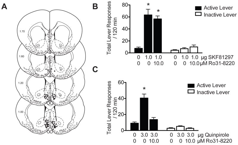
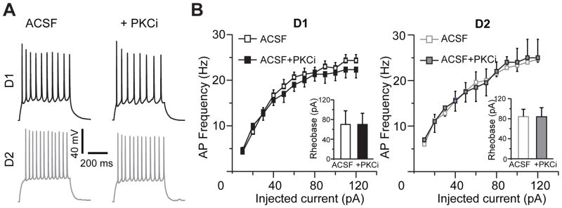
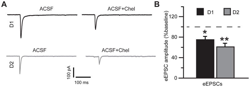
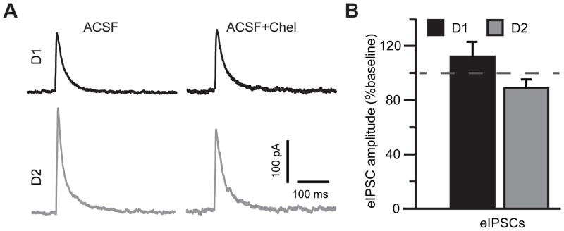
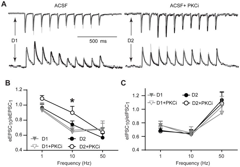
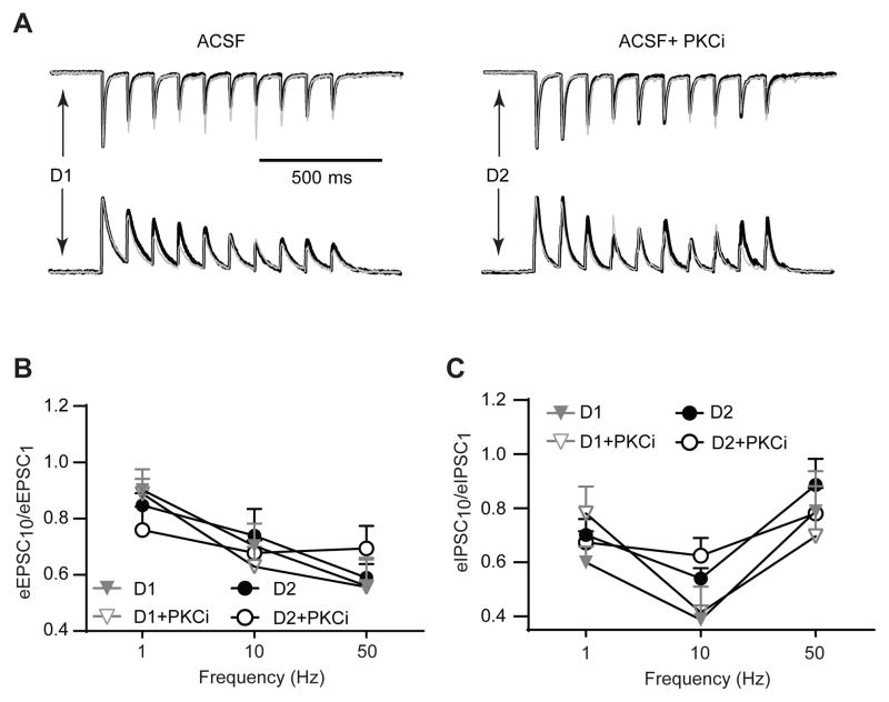
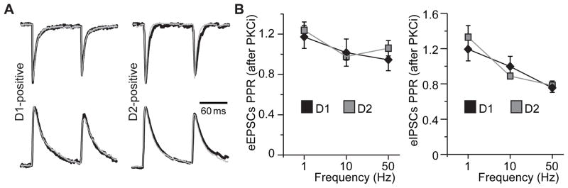
Stimulation of D1-like dopamine receptors (D1DRs) or D2-like dopamine receptors (D2DRs) in the nucleus accumbens (NAc) shell reinstates cocaine seeking in rats, an animal model of relapse. D2DRs and D1DRs activate protein kinase C (PKC) and recent studies indicate that activation of PKC in the NAc plays an important role in the reinstatement of drug seeking induced by a systemic cocaine priming injection. In the present study, pharmacological inhibition of PKC in the NAc shell attenuated cocaine seeking induced by intra-accumbens shell microinjection of a D2DR agonist, but not a D1DR agonist. D1DRs and D2DRs are primarily expressed on different accumbens medium spiny (MSN) neurons. Neuronal signaling and activity were assessed in these two populations of NAc neurons with transgenic mice expressing fluorescent labels under the control of D1DR and D2DR promoters. Following the extinction of cocaine self-administration, bath application of a PKC inhibitor produced similar effects on single evoked excitatory and inhibitory post-synaptic currents in D1DR- and D2DR-positive MSNs in the NAc shell. However, inhibition of PKC preferentially improved the ability of excitatory, but not inhibitory, synapses to sustain responding to brief train of stimuli specifically in D2DR-positive MSNs. This effect did not appear to involve modulation of presynaptic release mechanisms. Taken together, these findings indicate that the reinstatement of cocaine seeking is at least partially due to D2DR-dependent increases in PKC signaling in the NAc shell, which reduce excitatory synaptic efficacy in D2DR-expressing MSNs.

Keywords: Addiction; Dopamine; EPSCs; Electrophysiology; IPSCs; Relapse; Self-administration; Striatum.


Conflict of interest statement

Conflict of Interest

The authors declare no potential conflict of interest relating to this study.

Figures

Figure 1
Figure 1. Microinjection of a PKC inhibitor into the NAc shell blocks D2DR, but not D1DR, agonist-induced reinstatement of cocaine seeking
(A) Coronal sections depicting microinjection sites, as indicated by closed circles, targeting the medial NAc shell in the dopamine receptor agonist experiments. Numbers on the left side of the coronal sections denote distance from bregma in the anteroposterior direction. (B) Total number of lever responses during the reinstatement test session following intra-accumbens shell administration of vehicle, 1.0 μg SKF-81297 or 10.0 μM Ro31-8220 + 1.0 μg SKF-81297. There was a significant increase in active lever responding in animals treated with 1.0 μg SKF-81297 or 10.0 μM Ro 31-8220 + 1.0 μg SKF-81297 when compared to vehicle treated controls (Tukey’s HSD, *p<0.05). (C) Total lever responses following intra-accumbens shell infusion of vehicle, 3.0 μg quinpirole or 10.0 μM Ro 31-8220 + 3.0 μg quinpirole. There was a significant increase in active lever responding in animals treated with 3.0 μg quinpirole when compared to animals treated with vehicle or 10.0 μM Ro 31-8220 + 3.0 μg quinpirole (Tukey’s HSD, *p<0.05). No significant differences in inactive lever responding were observed in either the SKF-81297 or quinpirole experiments.
Figure 2
Figure 2. Action potential firing in NAc shell D1DR- and D2DR-positive MSNs is not affected by PKC inhibition
(A) Representative traces of action potentials elicited from the same D1DR- and D2DR-positive neuron in the NAc shell before and after PKC blockade. Responses to a current step of 60 pA above rheobase are shown in each cell. (B) The firing rate vs. input (f-I) curves illustrate minimal or no effect of PKC inhibition in D1DR-positive (left) or D2DR-positive (right) MSNs (n=5–7 cells from 2 animals). Insets, PKC blockade is likewise without effect on rheobase current in both cell types. PKCi, PKC inhibitor (chelerythrine (10 μM) or Ro 31-8220 (10 μM)).
Figure 3
Figure 3. PKC inhibition suppresses single stimulus eEPSCs in both D1DR- and D2DR-positive NAc shell MSNs
(A) eEPSC average traces from the same D1DR- and D2DR-positive cell in the absence and in the presence of PKC blockade. Stimulus artifacts were removed for illustration. (B) Peak amplitude of eEPSCs in D1DR- and D2DR-positive MSNs in the presence of PKC blockade is plotted as a percent of response recorded prior to PKC antagonist application. Note similar inhibition of eEPSC amplitude in both cell types (paired Student’s t-test, *, p<0.05; **, p<0.01; n=8–9 cells from 3 animals).
Figure 4
Figure 4. GABAA receptor-mediated single stimulus eIPSCs are not markedly affected by PKC inhibition
(A) eIPSC average traces from the same D1DR- and D2DR-positive cell in the absence and in the presence of PKC blockade. Stimulus artifacts were removed for illustration. (B) No significant differences in eIPSCs amplitude were observed in response to PKC antagonist application. Note, however, a trend toward PKCi-induced increase in D1DR-positive MSNs and PKC-induced decrease in D2DR-positive MSNs (n=8 cells from 3 animals).
Figure 5
Figure 5. Trains of stimuli differentially modulate synaptic excitation in NAc shell D1DR- and D2DR-positive MSNs following PKC blockade
(A) Top, representative eEPSCs elicited by a 1 second train of stimuli at 10 Hz in D1DR- and D2DR-positive MSNs. Thick, black traces recorded in the presence of PKC inhibition are overlaid onto thin gray traces recorded in the absence of PKC inhibition. PKC inhibition induces a consistent increase in eEPSC amplitude throughout the stimulus train seen in D2DR-, but not D1DR-positive MSNs. Stimulus artifacts were removed for illustration. Responses in each overlaid set of traces are normalized to the peak amplitude of the first pulse in the absence of PKC inhibition for ease of comparison. Bottom, no consistent response to PKC inhibition could be observed for eIPSCs in D1DR- and D2DR-positive MSNs. (B) An activity-dependent effect on eEPSC trains is expressed as a ratio of the last (eEPSC10) to first (eEPSC1) current response at 3 frequencies of stimulation. The difference between D2DR+PKCi and D1DR+PKCi groups is significant (*, p<0.05, Tukey’s post-hoc; n=9–12 cells from 4–5 animals). (C) Same as (B), but for eIPSCs. No significant differences were found (n=8–12 cells from 3–5 animals).
Figure 6
Figure 6. Stimulus trains do not affect fidelity of synaptic signaling in the absence of extracellular dopamine
(A) Top, representative eEPSCs elicited by a 1 second train of stimuli at 10 Hz in D1DR- and D2DR-positive MSNs. Thick, black traces recorded in the presence of PKC inhibition are overlaid onto thin gray traces recorded in the absence of PKC inhibition. PKC inhibition has similar effects on synaptic responding in D1DR- and D2DR-positive MSNs. Stimulus artifacts were removed for illustration. Responses in each overlaid set of traces are normalized to the peak amplitude of the first pulse in the absence of PKC inhibition for ease of comparison. Bottom, eIPSC responses to PKC inhibition are also similar in D1DR- and D2DR-positive MSNs. (B) Ratios of the last (eEPSC10) to first (eEPSC1) current response are plotted at 3 frequencies of stimulation. There are no significant differences between any of the groups (n=9–12 cells from 6 animals). (C) Same as (B), but for eIPSCs. No significant differences were found between groups (n=10–12 cells from 6 animals).
Figure 7
Figure 7. PKC inhibition does not affect the paired-pulse ratio of excitatory and inhibitory PSCs in MSNs of the NAc shell
(A) eEPSCs (top traces) or eIPSCs (bottom traces) evoked by a 10 Hz paired-pulse stimulus. Overlays of traces in the absence (thick black traces) and in the presence (thin gray traces) of PKC inhibition are pictured. Responses in each overlaid set of traces are normalized to the peak amplitude of the first pulse in the absence of PKC inhibition for ease of comparison. Stimulus artifacts were removed for illustration. (B) Paired-pulse ratios of eEPSCs (left) and eIPSCs (right) after PKC inhibition are plotted for D1DR- and D2DR-positive MSNs at indicated paired-pulse frequencies. No differences were found between any of the groups (n=8–9 cells from 3 animals).

References

    1. Albin RL, Young AB, Penney JB. The functional anatomy of disorders of the basal ganglia. Trends Neurosci. 1995;18:63–64. - PubMed
    1. Anderson SM, Bari AA, Pierce RC. Administration of the D1-like dopamine receptor antagonist SCH-23390 into the medial nucleus accumbens shell attenuates cocaine priming-induced reinstatement of drug-seeking behavior in rats. Psychopharmacology (Berl) 2003;168:132–138. - PubMed
    1. Anderson SM, Famous KR, Sadri-Vakili G, Kumaresan V, Schmidt HD, Bass CE, Terwilliger EF, Cha JH, Pierce RC. CaMKII: a biochemical bridge linking accumbens dopamine and glutamate systems in cocaine seeking. Nat Neurosci. 2008;11:344–353. - PubMed
    1. Anderson SM, Pierce RC. Cocaine-induced alterations in dopamine receptor signaling: implications for reinforcement and reinstatement. Pharmacol Ther. 2005;106:389–403. - PubMed
    1. Anderson SM, Schmidt HD, Pierce RC. Administration of the D2 dopamine receptor antagonist sulpiride into the shell, but not the core, of the nucleus accumbens attenuates cocaine priming-induced reinstatement of drug seeking. Neuropsychopharmacology. 2006;31:1452–1461. - PubMed

Publication types

MeSH terms