Review
. 2015 Aug;40(6):1788-99.
doi: 10.1007/s00261-015-0353-8.

Recent advances in image-guided targeted prostate biopsy

Affiliations
Review

Recent advances in image-guided targeted prostate biopsy

Anna M Brown et al. Abdom Imaging. 2015 Aug.

Abstract

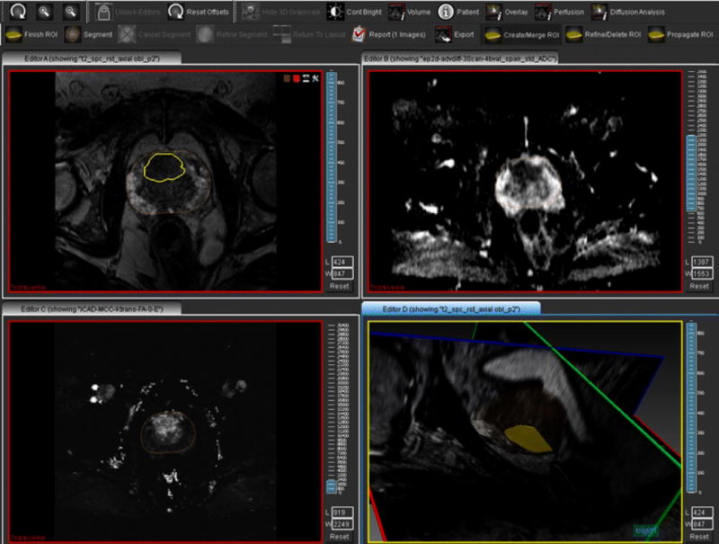
Prostate cancer is a common malignancy in the United States that results in over 30,000 deaths per year. The current state of prostate cancer diagnosis, based on PSA screening and sextant biopsy, has been criticized for both overdiagnosis of low-grade tumors and underdiagnosis of clinically significant prostate cancers (Gleason score ≥7). Recently, image guidance has been added to perform targeted biopsies of lesions detected on multi-parametric magnetic resonance imaging (mpMRI) scans. These methods have improved the ability to detect clinically significant cancer, while reducing the diagnosis of low-grade tumors. Several approaches have been explored to improve the accuracy of image-guided targeted prostate biopsy, including in-bore MRI-guided, cognitive fusion, and MRI/transrectal ultrasound fusion-guided biopsy. This review will examine recent advances in these image-guided targeted prostate biopsy techniques.


Figures

Fig. 1
Fig. 1
A 62 year old male with serum PSA of 23.85 ng/dL and previous TRUS-guided prostate biopsy showing Gleason 6 disease was followed by four subsequent TRUS-guided biopsies that were all negative despite substantially rising PSA levels. Axial T2-weighted MR image shows a hypointense area in the right mid base anterior peripheral zone also affecting the adjacent transition zone (a). The apparent diffusion coefficient (ADC) map shows a corresponding hypointense area (b), and the high b=2000 diffusion weighted MR image demonstrates hyperintensity (c). The dynamic contrast enhancement (DCE) sequence indicates early strong enhancement within the lesion as well (d)
Fig. 2
Fig. 2. Three different approaches for fusing prostate MRI with ultrasound
The use of electromagnetic tracking on a freehand transrectal ultrasound (TRUS) probe is found in three platforms, the UroNav, HI-RVS, and Virtual Navigator systems. Two of the platforms, Artemis and Biopsee, utilize an articulated approach with a mechanical stepper used to mount the ultrasound probe and embedded with encoders relating the position of the probe to the biopsy platform to aid with fusion. One other system, the Urostation, employs an image-based registration algorithm in which TRUS images are fused to the MRI to determine the appropriate biopsy location, and then TRUS is performed following biopsy sample acquisition to verify placement of the biopsy needle. Whereas the first two approaches (electromagnetic tracking and articulated) allow for real-time prospective targeting of suspicious areas, the TRUS-TRUS registration method only allows for retrospectively assessing the accuracy of the biopsy needle location. All of these systems enable operators to target lesions pre-identified on MRI in the realtime ultrasound suite in a more precise fashion than cognitive fusion, since images are either overlaid or deformably shaped to match contours from both modalities. Permission was obtained from InVivo, Eigen, and Koelis as indicated for use of their representative images
Fig. 3
Fig. 3
TRUS/MRI fusion guided biopsy procedure snapshot of a 62 year old male with serum PSA of 23.85 ng/dL (same patient in Fig. 2). The patient underwent MRI/TRUS fusion-guided biopsy using the UroNav platform. This figure shows a screen capture of the operator’s view during the biopsy procedure. The ultrasound and MRI scans have been co-registered elastically. The operator can freely rotate the ultrasound probe, and this will change the view of both the ultrasound image (superior) and the previously acquired MRI (inferior). The red dot with concentric green circle identifies the target lesion, which this operator has correctly ‘pierced’ with the biopsy needle. The location of the biopsy needle is shown by the yellow line, which can be documented for later retrieval on the UroNav system. Currently the routine prostate biopsy procedure for patients at the National Cancer Institute includes both conventional TRUS-guided biopsy and targeted biopsy performed at the same time. On histopathology, this patient was found to have high-grade Gleason 4+4=8 prostate adenocarcinoma (75% and 40% in 2 targeted cores) in this lesion. Only the targeted biopsy cores were positive for this patient, while all of the TRUS-guided biopsy cores showed benign prostatic tissue
Fig. 4
Fig. 4
Demonstration of the biopsy planning process for the Artemis system. The lesion is outlined in yellow at top left on the T2-weighted sequence and corresponds with the hypointense area on the ADC map shown at top right. The whole prostate outline is shown on the DCE image at bottom left, which demonstrates enhancement in the anterior region where the lesion is located. The bottom right panel shows a sagittal view of the MRI with lesion outlined
Fig. 5
Fig. 5
Longitudinal view of the output of the Artemis system following prostate biopsy, with the target lesion volumes shaded in gray and lines representing biopsy needle locations. The blue and purple dots represent the center of the biopsy needle where the cores were taken. Additional cores have been taken for the systematic biopsy locations, with the green dots (1–12) representing a template for the systematic biopsy sites

References

    1. What are the key statistics about prostate cancer? 2013:1. www.cancer.org.
    1. Sindhwani P, Wilson CM. Prostatitis and serum prostate-specific antigen. Curr Prostate Rep. 2005;3:81–86. doi: 10.1007/s11918-005-0020-6. - DOI - PubMed
    1. Bouraoui Y, Ricote M, García-Tuñón I, et al. Pro-inflammatory cytokines and prostate-specific antigen in hyperplasia and human prostate cancer. Cancer Detect Prev. 2008;32:23–32. doi: 10.1016/j.cdp.2008.02.007. - DOI - PubMed
    1. Lazzeri M, Abrate A, Lughezzani G, et al. Relationship of chronic histologic prostatic inflammation in biopsy specimens with serum isoform [−2]proPSA (p2PSA), %p2PSA, and prostate health index in men with a total prostate-specific antigen of 4–10 ng/ml and normal digital rectal examination. Urology. 2014;83:606–12. doi: 10.1016/j.urology.2013.10.016. - DOI - PubMed
    1. Hedelin H, Johansson N, Ströberg P. Relationship between benign prostatic hyperplasia and lower urinary tract symptoms and correlation between prostate volume and serum prostate-specific antigen in clinical routine. Scand J Urol Nephrol. 2005;39:154–9. doi: 10.1080/00365590510007685. - DOI - PubMed

Publication types