Review
. 2015 Mar 7;15(5):1230-49.
doi: 10.1039/c4lc01246a.

Microfluidic cell sorting: a review of the advances in the separation of cells from debulking to rare cell isolation

Affiliations
Review

Microfluidic cell sorting: a review of the advances in the separation of cells from debulking to rare cell isolation

C Wyatt Shields 4th et al. Lab Chip. .

Abstract

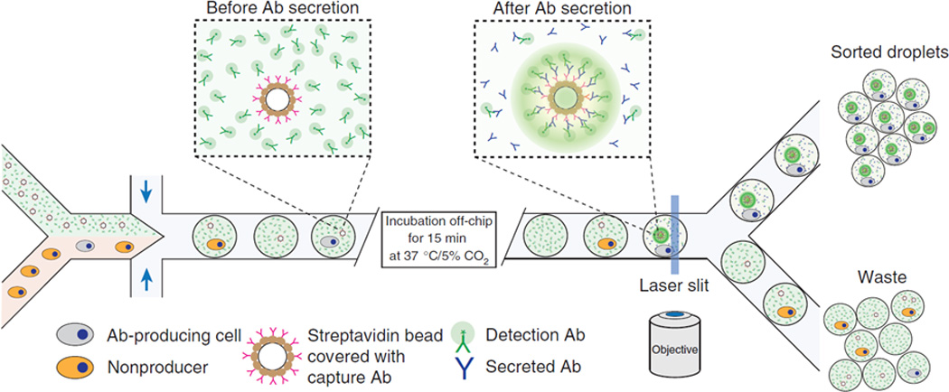
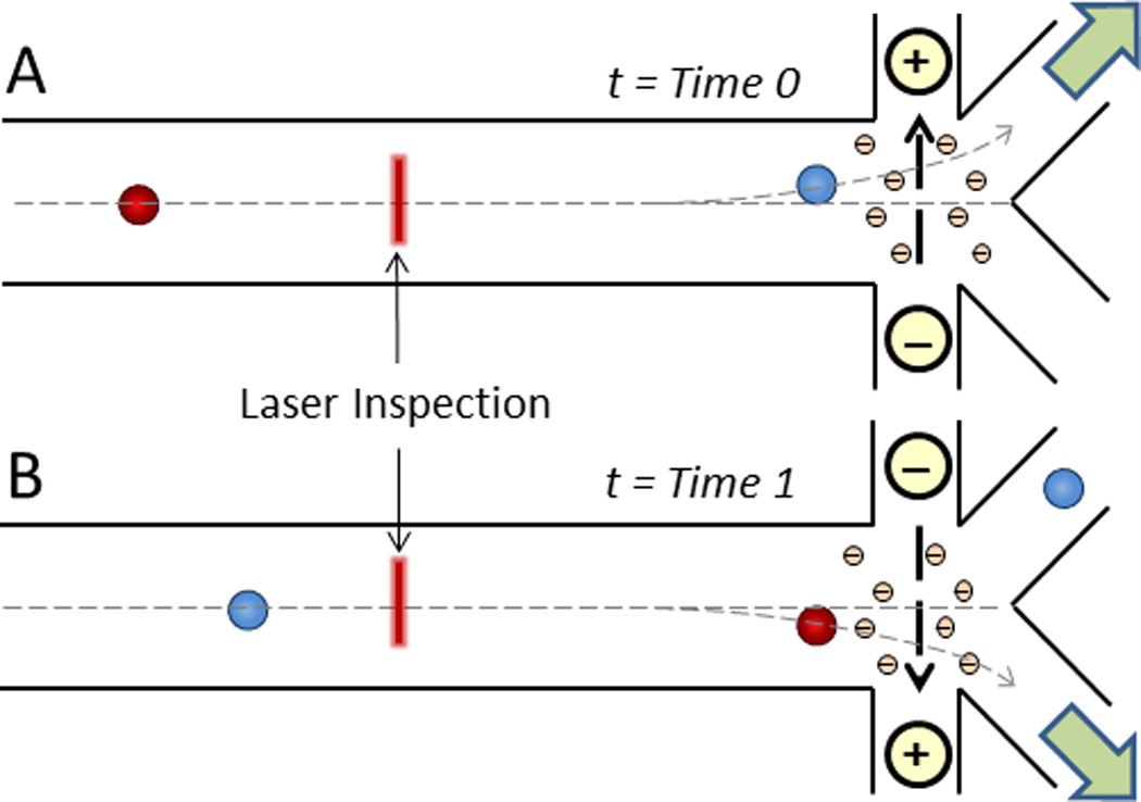
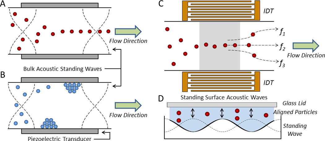
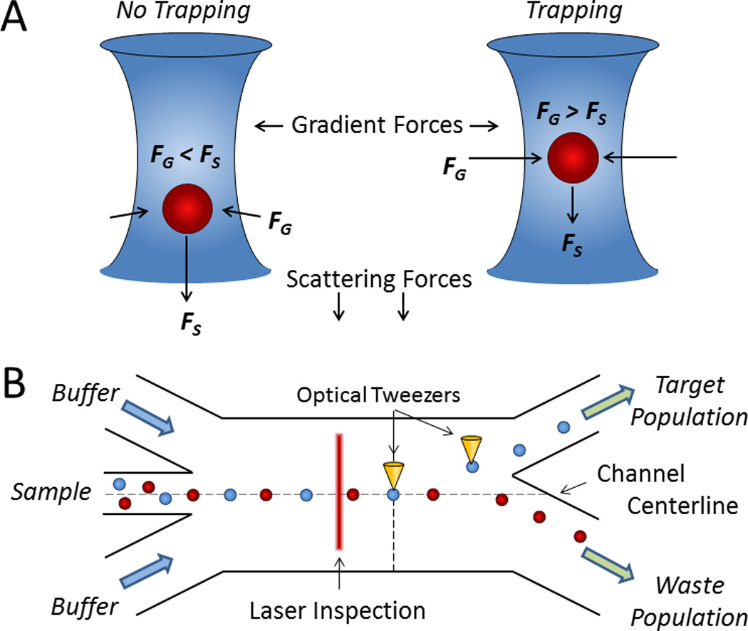
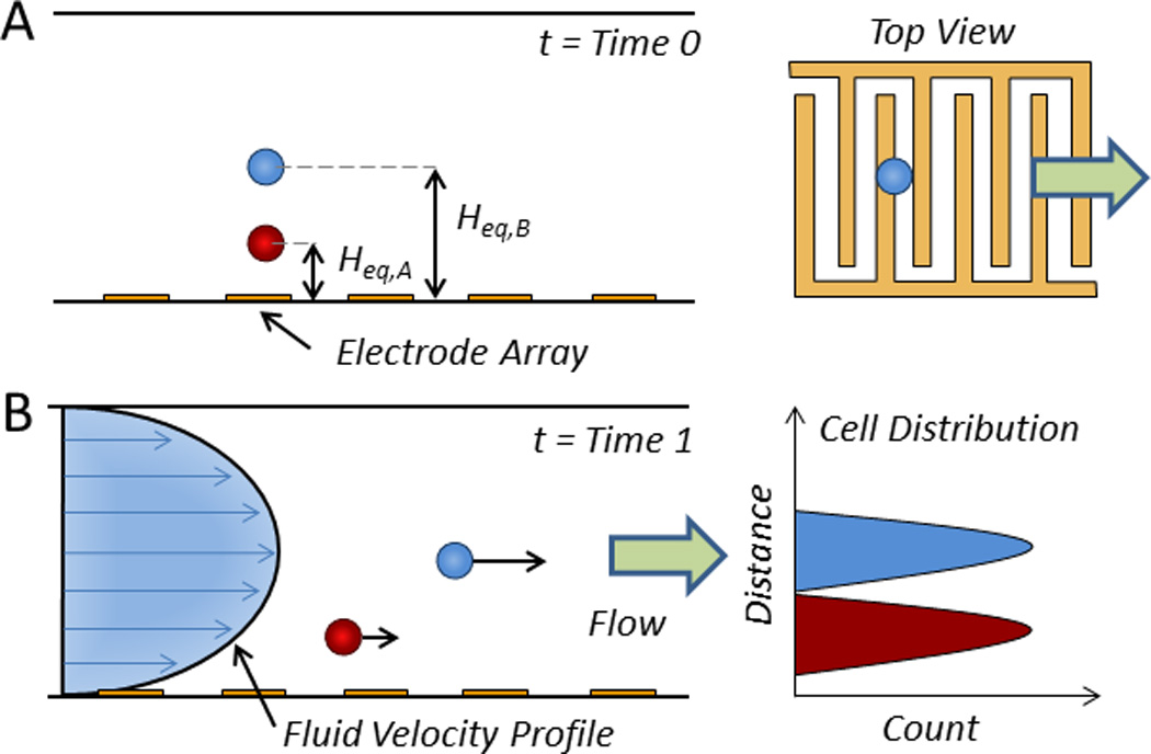
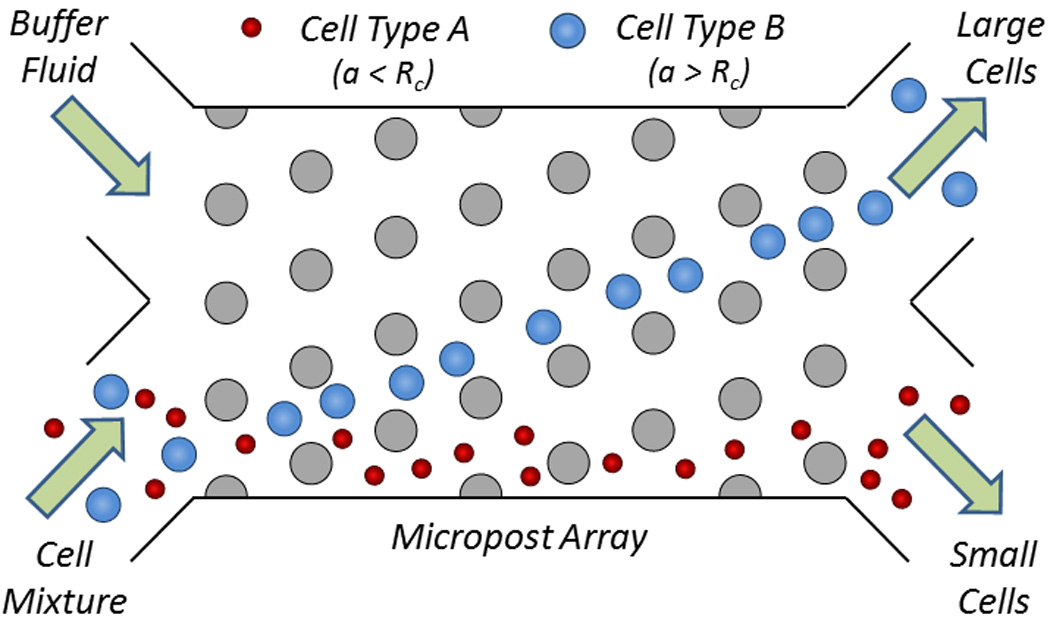
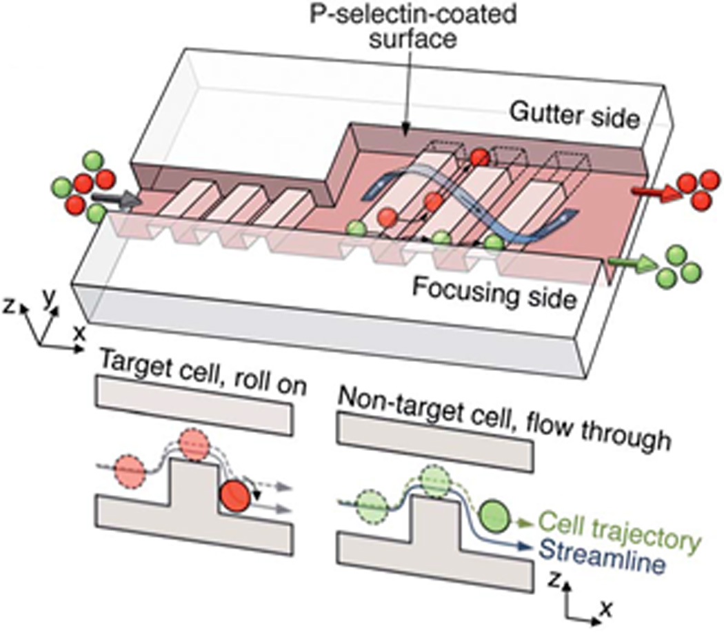
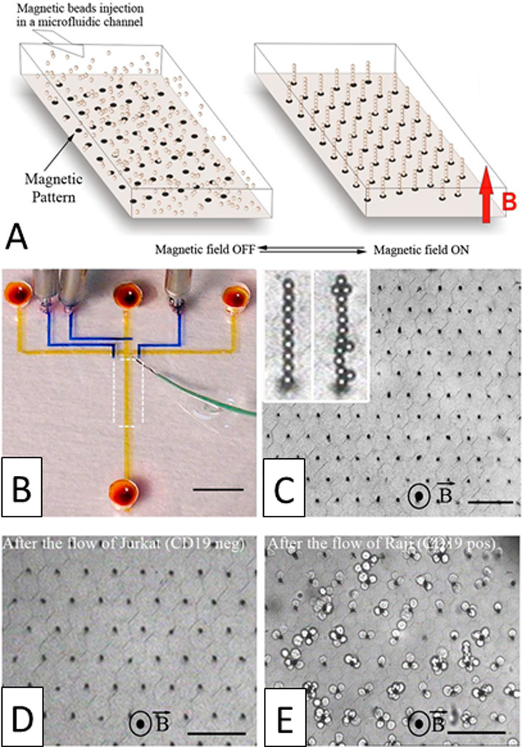
Accurate and high throughput cell sorting is a critical enabling technology in molecular and cellular biology, biotechnology, and medicine. While conventional methods can provide high efficiency sorting in short timescales, advances in microfluidics have enabled the realization of miniaturized devices offering similar capabilities that exploit a variety of physical principles. We classify these technologies as either active or passive. Active systems generally use external fields (e.g., acoustic, electric, magnetic, and optical) to impose forces to displace cells for sorting, whereas passive systems use inertial forces, filters, and adhesion mechanisms to purify cell populations. Cell sorting on microchips provides numerous advantages over conventional methods by reducing the size of necessary equipment, eliminating potentially biohazardous aerosols, and simplifying the complex protocols commonly associated with cell sorting. Additionally, microchip devices are well suited for parallelization, enabling complete lab-on-a-chip devices for cellular isolation, analysis, and experimental processing. In this review, we examine the breadth of microfluidic cell sorting technologies, while focusing on those that offer the greatest potential for translation into clinical and industrial practice and that offer multiple, useful functions. We organize these sorting technologies by the type of cell preparation required (i.e., fluorescent label-based sorting, bead-based sorting, and label-free sorting) as well as by the physical principles underlying each sorting mechanism.


Figures

Fig. 1
Fig. 1
Droplet-based microreactor and cell sorter using DEP. Cells expressing and secreting a target antibody (depicted in grey) and cells not expressing a target antibody (depicted in orange) are encapsulated in droplets with a fluorescent detection antibody, incubated off-chip to permit the production of secretion antibodies, and sorted according to the increased fluorescence signal from the localized packing of detection antibodies on the surface of the bead covered with capture and secretion antibodies. Reprinted with permission from Mazutis et al Copyright 2013 Nature Publishing Group.
Fig. 2
Fig. 2
Direct current (DC) electroosmotic cell sorting. Following laser inspection and cell identification, solvated negative ions in the counterionic layer along the positively charged microchannel floor migrate to the oppositely charged electrode, thereby dragging the surrounding liquid for cell transport to either A) the first outlet (t = Time 0) or B) the second outlet (t = Time 1).
Fig. 3
Fig. 3
Acoustofluidic manipulation of cells and particles. In a bulk acoustic standing wave, objects with a A) positive and B) negative ϕ migrate to the pressure node(s) and antinodes, respectively. C) In a SSAW device, interdigital transducers (IDTs) focus cells along well-defined streamlines according to the driving frequencies of the IDTs (e.g., f1, f2, and f3) for sorting across multiple outlets. D) The cross section of a SSAW device containing four pressure nodes.
Fig. 4
Fig. 4
Cell sorting by optical force switching. A) When scattering forces (FS) exceed gradient forces (FG) from a focused laser beam, cells are deflected (left); however, when FG exceeds FS, cells are optically trapped (right). B) A hydrodynamically focused stream of cells is aligned toward the waste outlet whereupon cells of interest detected by laser inspection are captured and displaced by optical tweezers for sorting.
Fig. 5
Fig. 5
Electromechanical T-switch for cell sorting. A microfabricated cantilever beam reversibly shifts from A) a ‘down’ position (t = Time 0) to B) an ‘up’ position (t = Time 1) when a corresponding pair of electrodes is activated to generate bubbles via isothermal electrolysis, which in turn exerts a mechanical force on the T-switch to redirect fluid flow.
Fig. 6
Fig. 6
Magnetic isolation of CTCs in microfluidic devices. A) Magnetic beads conjugated with anti-EpCAM are shown to capture and isolate CTCs under free flow in a magnetic field. Reprinted with permission from Hoshino et al Copyright 2011 Royal Society of Chemistry. B) Magnetically labeled CTCs and leukocytes are filtered from blood via deterministic lateral displacement (see Fig. 12), focused via inertial focusing (see Fig. 10A), and sorted in a magnetic field. Reprinted with permission from Ozkumar et al Copyright 2013 Science Translational Medicine.
Fig. 7
Fig. 7
Acoustic separation of cells using elastomeric particles. A) Elastomeric particles and cells focus to the antinodes and node(s) of an acoustic standing wave, respectively. B) When elastomeric particles bind to target cells, those complexes displace to the pressure antinodes for separation from non-target cells. Adapted from Shields IV et al 2014 American Chemical Society.
Fig. 8
Fig. 8
Multi-target cell sorting using DEP and magnetic trapping. A) Target Cell A is captured with polystyrene beads depicted in green and Target Cell B is captured with superparamagnetic beads depicted in red. B) Target Cell A is removed from non-target cells (depicted in blue) by dielectrophoretic forces and Target Cell B is removed from non-target cells by magnetic trapping. Reprinted with permission from Kim et al Copyright 2009 Royal Society of Chemistry.
Fig. 9
Fig. 9
Sorting by nDEP-assisted field-flow fractionation. A) An array of electrodes displaces cells to equilibrium positions above the floor of a microfluidic channel according to their type (t = Time 0) and B) sorts those cells by propelling them through the channel at rates according to their distance from the wall (t = Time 1).
Fig. 10
Fig. 10
Inertial microfluidics for cell sorting in curved microchannels. A) A serpentine microfluidic channel can focus cells into a single streamline. B) A spiral microfluidic channel can sort cells by size (IW and OW indicate the inner wall and outer wall, respectively). C) The cross section of the microfluidic channel showing the balance between lift forces (FL) and Dean drag forces (FD). Reprinted with permission from Kuntaegowdanahalli et al Copyright 2009 Royal Society of Chemistry.
Fig. 11
Fig. 11
Cell sorting by pinched flow fractionation. A) In the pinched segment, cells are first pushed against the wall, and then separated by size upon broadening of the microfluidic channel. B) Cells are aligned in the pinched segment of the channel and follow separate streamlines for sorting by size after exiting the pinched segment. Reprinted with permission from Yamada et al Copyright 2004 American Chemical Society.
Fig. 12
Fig. 12
Cell sorting by deterministic lateral displacement. Large cells (depicted in blue) migrate away from the small cells (depicted in red) in the initial streamline due to the engineered size and spacing of the microposts in the microfluidic channel.
Fig. 13
Fig. 13
Hydrophoretic cell focusing and sorting. A) A simplified free-body diagram of cells with a low density (shown in blue) separating from cells with a high density (shown in red). The black arrows pointing upwards represent buoyancy forces and the black arrows pointing downwards represent settling forces. B) A top view of the microfluidic channel with herringbone grooves in the ceiling to guide the focusing and separation of cells by density.
Fig. 14
Fig. 14
Micropatterned ratchets for isolating cells by size and deformability. A) A design of microratchet funnels for fractionating cell populations (dimensions in µm). B) The operation area of the device whereby cells (1, left) enter the chip, (2) reversibly flow through the ratchets for separation by size, and (1, right) exit the chip. C) Schematic of a size exclusion filtration device with various inlets, outlets, and valves (V1-V6). Reprinted with permission from McFaul et al Copyright 2012 Royal Society of Chemistry.
Fig. 15
Fig. 15
Microfluidic filtration mechanisms. Schematic of a A) weir filter, B) pillar filter, and C) cross-flow filter to separate smaller cells (depicted in red) from larger cells (depicted in blue).
Fig. 16
Fig. 16
Cell sorting by hydrodynamic filtration. A) Cells are injected into the microfluidic device and are pushed toward the outlets. B) Small cells exit out of the proximal branches whereas C) large cells exit out of the distal branches. Reprinted with permission from Yamada et al Copyright 2007 Springer.
Fig. 17
Fig. 17
Cell sorting by deterministic cell rolling. Target cells (red) interact with the surface, roll across the ridges, and laterally displace toward the gutter side whereas non-target cells (green) flow over the ridges, not interacting with the surface, and exit on the focusing side. Reprinted with permission from Choi et al Copyright 2012 Royal Society of Chemistry.
Fig. 18
Fig. 18
Magnetic self-assembly of biofunctional magnetic beads for isolating rare cells. A) Schematic of a hexagonal array of magnetic ink (left) can guide the self-assembly of magnetic beads conjugated with anti-CD19 mAb in the presence of a vertical magnetic field (right). B) Photograph of the microfluidic device. Optical micrographs of the columns after C) the assembly of magnetic beads, D) the passage of 1,000 Jurkat cells (CD19 negative), and E) the passage of 400 Raji cells (CD19 positive) (scale bar: 80 µm). Reprinted with permission from Saliba et al Copyright 2010 Proceedings of the National Academy of Sciences of the USA.

References

    1. Tomlinson MJ, Tomlinson S, Yang XB, Kirkham J. Journal of tissue engineering. 2013;4:2041731412472690. - PMC - PubMed
    1. Armstrong AJ, Marengo MS, Oltean S, Kemeny G, Bitting RL, Turnbull JD, Herold CI, Marcom PK, George DJ, Garcia-Blanco MA. Molecular cancer research : MCR. 2011;9:997–1007. - PMC - PubMed
    1. Wognum AW, Eaves AC, Thomas TE. Archives of medical research. 2003;34:461–475. - PubMed
    1. Bischoff F, Marquez-Do D, Martinez D, Dang D, Horne C, Lewis D, Simpson L. Clin Genet. 2003;63:483–489. - PubMed
    1. Chen Y, Li P, Huang PH, Xie Y, Mai JD, Wang L, Nguyen NT, Huang TJ. Lab Chip. 2014;14:626–645. - PMC - PubMed

Publication types

Substances