Review
. 2015 Jan-Feb;16(1):99-113.
doi: 10.3348/kjr.2015.16.1.99. Epub 2015 Jan 9.

Imaging findings of common benign renal tumors in the era of small renal masses: differential diagnosis from small renal cell carcinoma: current status and future perspectives

Affiliations
Review

Imaging findings of common benign renal tumors in the era of small renal masses: differential diagnosis from small renal cell carcinoma: current status and future perspectives

Sungmin Woo et al. Korean J Radiol. 2015 Jan-Feb.

Abstract

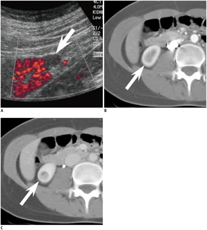
The prevalence of small renal masses (SRM) has risen, paralleling the increased usage of cross-sectional imaging. A large proportion of these SRMs are not malignant, and do not require invasive treatment such as nephrectomy. Therefore, differentiation between early renal cell carcinoma (RCC) and benign SRM is critical to achieve proper management. This article reviews the radiological features of benign SRMs, with focus on two of the most common benign entities, angiomyolipoma and oncocytoma, in terms of their common imaging findings and differential features from RCC. Furthermore, the role of percutaneous biopsy is discussed as imaging is yet imperfect, therefore necessitating biopsy in certain circumstances to confirm the benignity of SRMs.

Keywords: Angiomyolipoma; Oncocytoma; Renal cell carcinoma; Small renal mass.


Figures

Fig. 1
Fig. 1
Comparison of relative echogenicity between small renal cell carcinoma (RCC) and angiomyolipoma (AML) with minimal fat. A. Mass (arrow) in right kidney lower pole in 43-year-old woman shows high echogenicity but less than that of sinus fat. Relative echogenicity ([echogenicity of mass - echogenicity of renal cortex] / [echogenicity of sinus fat - echogenicity of renal cortex]) was calculated as 0.63. Upon surgery, mass was confirmed as clear cell RCC. B. Mass (arrow) in left kidney interpolar in 38-year-old woman shows high echogenicity, even higher than that of sinus fat. Relative echogenicity was measured as 2.10. Mass was confirmed as AML with minimal fat at surgery.
Fig. 2
Fig. 2
year-old man with small angiomyolipoma with minimal fat in right kidney demonstrating typical CT and MR findings. A. Axial precontrast CT scan reveals oval shaped 1.8-cm sized hyperdense mass (arrow) in right kidney upper pole. At region of interest measurement, attenuation of mass was 51 Hounsfield units (HU) while renal parenchyma was measured as 39 HU. B, C. Axial corticomedullary phase (B) and early excretory phase (C) CT scan shows that mass (arrow) is less enhanced compared with renal parenchyma. D. Renal mass (arrow) demonstrates low signal intensity (SI) compared with renal parenchyma on coronal T2-weighted image. E, F. On coronal chemical shift MR imaging, suspicious focus (arrow) of SI drop from in phase (E) to opposed phase (F) is noted. G-I. Coronal contrast-enhanced MRI shows that mass (arrow) is less enhanced than background renal parenchyma.
Fig. 3
Fig. 3
53-year-old man with small angiomyolipoma with minimal fat showing ice-cream cone appearance. A. Small enhancing renal mass in left kidney anterior aspect is depicted on axial corticomedullary phase CT. Mass is composed of two portions, exophytic portion (arrow) with rounded appearance and intraparenchymal component (arrowhead) with wedge or triangular shape, resembling ice-cream cone. B. At sagittal early excretory phase CT, ice-cream cone appearance constituted with exophytic (arrow) and endophytic portions (arrowhead) of renal mass is again well demonstrated. Mass was diagnosed as angiomyolipoma with minimal fat at surgery.
Fig. 4
Fig. 4
44-year-old woman with small oncocytoma demonstrating segmental enhancement inversion. A. On coronal corticomedullary phase CT, renal mass at right kidney upper pole can be segmented into two areas. Crescent-shaped area at right aspect (white arrow) is more enhanced compared with relatively round shaped portion (black arrow) with heterogeneous enhancement at left aspect. Note adjacent medulla (arrowhead) at lateral aspect of renal mass which is less enhanced compared with well enhancing portion of renal mass. B. On coronal early excretory phase CT, enhancement degree of aforementioned two segments of renal mass is reversed. While previously more enhanced right crescent-shaped area (white arrow) is now less enhanced, round portion at left aspect (black arrow) shows marked enhancement, consistent with segmental enhancement inversion. Note that corticomedullary phase hypodense area and early excretory phase hyperattenuating area (arrowhead) are not part of mass but adjacent medullary tissue.
Fig. 5
Fig. 5
33-year-old woman with small metanephric adenoma. A. Mass at right kidney upper pole is slightly more hyperdense (arrow) than renal parenchyma at axial precontrast CT. B, C. Mass (arrow) is poorly enhancing compared with renal parenchyma at axial corticomedullary phase (B) and early excretory phase (C). Mass was confirmed as metanephric adenoma upon surgical resection.
Fig. 6
Fig. 6
43-year-old woman with small renal leiomyoma. A. Renal mass (arrow) is located at right kidney capsular area abutting right hemiliver. On axial precontrast CT scan, mass is hyperdense in comparison with renal parenchyma. B, C. Axial corticomedullary (B) and early excretory phase (C) CT reveals that renal mass (arrow) is homogeneously enhanced. Renal mass was diagnosed as leiomyoma after surgery.
Fig. 7
Fig. 7
23-year-old woman with small juxtaglomerular cell tumor and underlying hypertension. A. Power Doppler ultrasound demonstrates renal mass (arrow) with poor vascularity. B, C. Axial corticomedullary (B) and nephrographic (C) CT shows same mass (arrow) without prominent enhancement in right kidney. At surgery, mass was confirmed as juxtaglomerular cell tumor.

References

    1. Silverman SG, Israel GM, Herts BR, Richie JP. Management of the incidental renal mass. Radiology. 2008;249:16–31. - PubMed
    1. Gill IS, Aron M, Gervais DA, Jewett MA. Clinical practice. Small renal mass. N Engl J Med. 2010;362:624–634. - PubMed
    1. Chow WH, Dong LM, Devesa SS. Epidemiology and risk factors for kidney cancer. Nat Rev Urol. 2010;7:245–257. - PMC - PubMed
    1. Reis LAG, Melbert D, Krapcho M, Stinchcomb DG, Howlader N, Horner MJ, et al. SEER Cancer Statistics Review, 1975-2007. Bethesda, MD: National Cancer Institute; 2008.
    1. Rioux-Leclercq N, Karakiewicz PI, Trinh QD, Ficarra V, Cindolo L, de la Taille A, et al. Prognostic ability of simplified nuclear grading of renal cell carcinoma. Cancer. 2007;109:868–874. - PubMed

MeSH terms