. 2014 Mar;3(1):1450003.
doi: 10.1142/S2047684114500031.

Computational conformational antimicrobial analysis developing mechanomolecular theory for polymer biomaterials in materials science and engineering

Affiliations

Computational conformational antimicrobial analysis developing mechanomolecular theory for polymer biomaterials in materials science and engineering

Richard C Petersen. Int J Comput Mater Sci Eng. 2014 Mar.

Abstract

Single-bond rotations or pyramidal inversions tend to either hide or expose relative energies that exist for atoms with nonbonding lone-pair electrons. Availability of lone-pair electrons depends on overall molecular electron distributions and differences in the immediate polarity of the surrounding pico/nanoenvironment. Stereochemistry three-dimensional aspects of molecules provide insight into conformations through single-bond rotations with associated lone-pair electrons on oxygen atoms in addition to pyramidal inversions with nitrogen atoms. When electrons are protected, potential energy is sheltered toward an energy minimum value to compatibilize molecularly with nonpolar environments. When electrons are exposed, maximum energy is available toward polar environment interactions. Computational conformational analysis software calculated energy profiles that exist during specific oxygen ether single-bond rotations with easy-to-visualize three-dimensional models for the trichlorinated bisaromatic ether triclosan antimicrobial polymer additive. As shown, fluctuating alternating bond rotations can produce complex interactions between molecules to provide entanglement strength for polymer toughness or alternatively disrupt weak secondary bonds of attraction to lower resin viscosity for new additive properties with nonpolar triclosan as a hydrophobic toughening/wetting agent. Further, bond rotations involving lone-pair electrons by a molecule at a nonpolar-hydrocarbon-membrane/polar-biologic-fluid interface might become sufficiently unstable to provide free mechanomolecular energies to disrupt weaker microbial membranes, for membrane transport of molecules into cells, provide cell signaling/recognition/defense and also generate enzyme mixing to speed reactions.

Keywords: Conformational analysis; bond rotation; inversion; lone-pair electrons; mechanomolecular; nonpolar; polar.


Figures

Fig. 1
Fig. 1
Two-dimensional (2D) molecular structure for triclosan showing center oxygen containing two lone-pair electrons and ether bonds between the two conjugated planar aromatic rings. Also, triclosan is trichlorinated and has one hydroxyl group on an aromatic ring in the ortho position.
Fig. 2
Fig. 2
Bacteria with no nucleus attach chromosomes to the cell membrane that subsequently invaginates inward between the two circular-like chromosomes with a septum during the rapid binary fission process.
Fig. 3
Fig. 3
The energy profile for triclosan is charted with bond rotation about the oxygen ether atom comparing the relative bond energies for bond angles between 20° to 90° rotation that gave an energy minimum at about 30° bond angle rotation.
Fig. 4
Fig. 4
Bond angle of approximately 30° rotation maintains hidden oxygen ether atom and lone-pair electrons with the dipole moment also concealed between aromatic rings corresponding to energy minimum observed in the charted results for Fig. 3.
Fig. 5
Fig. 5
90° bond rotation of both phenyl rings through oxygen ether bonds demonstrating how oxygen lone-pair electrons are completely exposed with dipole moment corresponding to the high bond energy value observed in the charted results for Fig. 3.
Fig. 6
Fig. 6
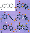
(a)–(d) Four identical conformations of triclosan at 90° oxygen ether bond rotation viewed from different angles for a stereoscopic perspective on polar access to both lone-pair electrons of the center ether oxygen atom with exposed dipole moment viewed from the same side as Fig. 1.
Fig. 7
Fig. 7
Triclosan oxygen ether bond rotations from 2D 0.0° rotation through to 180° in 3D: (a) Common 2D structure 0.0° ether bond rotation. (b) 3D structure 45.0° ether bond rotation. (c) 3D structure 90.0° ether bond rotation. (d) 3D structure 120.0° ether bond rotation. (e) 3D structure 150.0° ether bond rotation. (f) 3D structure 180.0° ether bond rotation with steric interaction between hydrogen atoms.
Fig. 8
Fig. 8
Increasing average flexural strength values are measured as triclosan is incorporated into the BisGMA/TEGDMA photocure resin up to 20wt.%.
Fig. 9
Fig. 9
Increases in average flexural strength is noted when adding triclosan into the photocure 84.5wt.% zirconia silicate particulate-filled composite up to 4.25wt.%.
Fig. 10
Fig. 10
A slight increase for average flexural strength is measured when adding 10 wt.% triclosan into the 35wt.% quartz fiber-reinforced particulate-filled photocure composite.
Fig. 11
Fig. 11
Increase in average flexural strength with 10 wt.% triclosan to chemical-cure acrylic formulation.
Fig. 12
Fig. 12
Condensing index shows increasing loss of paste consistency during compressive insertion of force gauge measurement tool that appears significantly exponential for the regression from 0.0wt.% to 15.31wt.% triclosan additions into the 84.5wt.% zirconia silicate particulate-filled composite.
Fig. 13
Fig. 13
Triclosan oxygen ether bond rotation of 30°is further influenced by intramolecular secondary hydrogen bonding from the hydroxyl group with the ether oxygen atom that connects both aromatic rings or the chlorine atom on the opposite phenyl ring to increase stability at the energy minimum position from any further rotation away from the initial 2D 0.0° rotation planar state shown by Fig. 1.
Fig. 14
Fig. 14
Chemical structure for triclosan methyl is a liquid at room temperature showing a methyl group replacing the hydrogen atom on the hydroxyl phenol group of triclosan that exists as a crystalline powder.
Fig. 15
Fig. 15
Low-energy minimum at 30°bond rotation is nonpolar and hydrophobic while the high-energy at 90°bond rotation is polar and hydrophilic. Changes in bond rotation between maximum and minimum relative energies are approximately 60°at −1.3kJ/mol.
Fig. 16
Fig. 16
Triclosan molecule oxygen ether bond rotations compatibilize well with the BisGMA polymer unit for aromatic rings and similar ether functions for single bonds that rotate to influence nonbonding mechanically-related entanglement to normally increase the strength of polymers and possibly toughness.
Fig. 17
Fig. 17
(a) Top structure BisGMA resin chain polymer unit has a viscosity of 700,000 cps related to hydroxyl groups that form hydrogen bonds. (b) Lower structure EBPADMA resin chain polymer unit without hydroxyl groups has a viscosity of just 775 cps that is almost three orders of magnitude lower than BisGMA.
Fig. 18
Fig. 18
Triclosan mechanomolecular energy disrupts hydrogen bonding between BisGMA resin chains thought responsible for the extremely high viscosity of 700,000 cps.
Fig. 19
Fig. 19
Molecular structure of triclocarban similar to bisaromatic triclosan but with diamide-style linkage instead of an ether linkage and no hydroxyl group.
Fig. 20
Fig. 20
Pyramidal inversion of nitrogen atom to expose lone-pair electrons for polar or hydrophilic media and alternatively protect the lone-pair electrons from nonpolar or hydrophobic media.
Fig. 21
Fig. 21
(a) Newman projection shows bond rotations for butane. (b) Relative energies during butane bond rotation. Bond rotations favor a minimum with methyl groups in butane as far away from one another as possible. The highest least stable conformation occurs when the methyl groups in butane are eclipsed directly in front of one another. Relative energies for a methyl group eclipsed with hydrogen require 16 kJ/mol and both methyl groups eclipsed 19 kJ/mol.
Fig. 22
Fig. 22
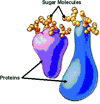
Basic globular protein or glycoprotein represented with sugar polysaccharide molecules attached (with Permission National Institutes of Health/Department of Health and Human Services).
Fig. 23
Fig. 23
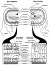
Comparison of the similarities for cell membrane envelopes between gram negative and gram positive bacteria. Triclosan molecules outside of the inner membrane side are shown for relative contrast of sizes to better appreciate the possible forces involved through the broad aromatic ring moment arms during oxygen ether bond rotation. Triclosan forces appear to accumulate in perpendicular alignment by aromatic pi–pi ring stacking and hydrogen bonding into the membrane near the phosphate head groups. Note the extra outer membrane for the gram negative cell (with permission from Salton and Kim [1996]).
Fig. 24
Fig. 24
Comparison between gram positive and gram negative bacterial strains for triclosan minimum inhibitory concentrations by agar incorporation testing [data from Ciba Specialty Chemicals, 2001].
Fig. 25
Fig. 25
Plasma cell membrane showing relationships for phospholipids, sugars, proteins and cholesterol. The eukaryote plasma cell membrane has some basic difference from the bacterial prokaryote cell membrane with cholesterol that prevents crystallization of lipid chains in addition to a comprehensive intracellular linkage with an extensive cytoskeleton not shown. For a comparative example relationship, triclosan molecules shown on the extracellular side of the lipid membrane tend to accumulate in membranes near the phosphate headgroups.
Fig. 26
Fig. 26
Eukaryote cells that include mammalians have a nucleus and extensive cytoskeleton protein fiber network that supports the cell membrane. (a) Drawing model emphasizes the nucleus that protects chromosomes and cytoskeleton network that supports the cell membrane. (b) Microscope image includes nucleus with microtubules of the cytoskeleton accentuated by special dyes (with permission from National Institutes of Health/Department of Health and Human Services).
Fig. 26
Fig. 26
Eukaryote cells that include mammalians have a nucleus and extensive cytoskeleton protein fiber network that supports the cell membrane. (a) Drawing model emphasizes the nucleus that protects chromosomes and cytoskeleton network that supports the cell membrane. (b) Microscope image includes nucleus with microtubules of the cytoskeleton accentuated by special dyes (with permission from National Institutes of Health/Department of Health and Human Services).

Similar articles

Cited by

References

    1. Akaki E, Mansur HS, Angelis LH, Castro BA, Valadão HF, Faria DB, Rezende FC. SEM/EDX and FTIR characterization of a dental resin cement with antibacterial agents incorporated. Key Eng. Mater. 2005;284–286:391–394.
    1. Alberts B, Johnson A, Lewis J, Raff M, Roberts K, Walters P. Molecular Biology of the Cell. 4th edn. New York: Garland Science; 2002.
    1. Anusavice KJ. Science of Dental Materials. 11th edn. St. Louis: Saunders Elsevier Science; 2003.
    1. Atkins PW. Physical Chemistry. 5th edn. New York: W. H. Freeman and Company; 1994.
    1. Australian Government, Department of Health and Ageing NICNAS. Priority Existing Chemical Assessment Report No. 30, Triclosan. Sydney, Australia: National Industrial Chemical Notification and Assessment Scheme; 2009. http://www.nicnas.gov.au/publications/car/pec/pec30/pec_30_full_report_p....

LinkOut - more resources