

Effects of defining realistic compositions of the ocular melanoma on proton therapy
- PMID: 25599060
- PMCID: PMC4289521
Effects of defining realistic compositions of the ocular melanoma on proton therapy
Abstract
Background: Recent studies in eye plaque brachytherapy have shown a considerable difference between the dosimetric results using water phantom and a model of human eye containing realistic materials. In spite of this fact, there is a lack of simulation studies based on such a model in proton therapy literatures. In the presented work, the effect of utilizing an eye model with ocular media on proton therapy is investigated using the MCNPX Monte Carlo Code.
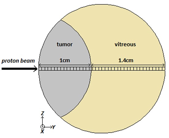
Methods: Two different eye models are proposed to study the effect of defining realistic materials on dose deposition due to utilizing pencil beam scanning (PBS) method for proton therapy of ocular melanoma. The first model is filled with water, and the second one contains the realistic materials of tumor and vitreous. Spread out Bragg peaks (SOBP) are created to cover a typical tumor volume. Moreover, isodose curves are figured in order to evaluate planar variations of absorbed dose in two models.
Results: The results show that the maximum delivered dose in ocular media is approximately 12-32% more than in water phantom. Also it is found that using the optimized weighted beams in water phantom leads to disturbance of uniformity of SOBP in ocular media.
Conclusion: Similar to the results reported in eye brachytherapy published papers, considering the ocular media in simulation studies leads to a more realistic assessment of sufficiency of the designed proton beam in tissue. This effect is of special importance in creating SOBP, as well as in delivered dose in the tumor boundaries in proton pencil beam scanning method.
Keywords: Dosimetry; MCNPX; Proton therapy; SOBP; Uveal melanoma.
Figures


References
-
- Stallard HB. Malignant melanoblastoma of the choroid. Bibl Ophthalmol. 1968;75:16–38. PubMed PMID: 5641363. - PubMed
-
- Damato B. Ocular tumours: Diagnosis and treatment. Oxford: Butterworth Heinemann; 2000. p. 250.
-
- Wilson RR. Radiological use of fast protons. Radiology. 1946;47(5):487–91. doi: 10.1148/47.5.487. PubMed PMID: 20274616. - PubMed
-
- Constable IJ, Roehler AM. Experimental ocular irradiation with accelerated protons. Invest Ophthalmol. 1974;13(4):280–7. PubMed PMID: 4206547. - PubMed
-
- Damato B, Kacperek A, Chopra M, Campbell IR, Errington RD. Proton beam radiotherapy of choroidal melanoma: the Liverpool-Clatterbridge experience. Int J Radiat Oncol Biol Phys. 2005;62(5):1405–11. doi: 10.1016/j.ijrobp.2005.01.016. PubMed PMID: 16029800. - PubMed
LinkOut - more resources
Full Text Sources