. 2014 Nov 1:1:261-283.
doi: 10.1146/annurev-virology-031413-085537.

IFITM-Family Proteins: The Cell's First Line of Antiviral Defense

Affiliations

IFITM-Family Proteins: The Cell's First Line of Antiviral Defense

Charles C Bailey et al. Annu Rev Virol. .

Abstract

Animal cells use a wide variety of mechanisms to slow or prevent replication of viruses. These mechanisms are usually mediated by antiviral proteins whose expression and activities can be constitutive but are frequently amplified by interferon induction. Among these interferon-stimulated proteins, members of the IFITM (interferon-induced transmembrane) family are unique because they prevent infection before a virus can traverse the lipid bilayer of the cell. At least three human IFITM proteins-IFITM1, IFITM2, and IFITM3-have antiviral activities. These activities limit infection in cultured cells by many viruses, including dengue virus, Ebola virus, influenza A virus, severe acute respiratory syndrome coronavirus, and West Nile virus. Murine Ifitm3 controls influenza A virus infection in vivo, and polymorphisms in human IFITM3 correlate with the severity of both seasonal and highly pathogenic avian influenza virus. Here we review the discovery and characterization of the IFITM proteins, describe the spectrum of their antiviral activities, and discuss potential mechanisms underlying these effects.

Keywords: Ebola virus; IFITM3; SARS coronavirus; dengue virus; flavivirus; influenza A virus; interferon; restriction factor; viral entry.


Figures
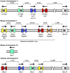
Figure 1
Figure 1
The immunity-related IFITM genes (IFITM1, IFITM2, and IFITM3), together with the osteoblast-restricted IFITM5, form a gene cluster in humans that is conserved in mice and chickens (25, 26). Alternative gene names are given in parentheses. Exons are depicted in color and introns in dark gray. Arrows indicate the direction of transcription. Mice possess two additional immunity-related Ifitm genes, Ifitm6 and Ifitm7 (19). Ifitm6 lies within the chromosome 7 Ifitm cluster, but Ifitm7 is a retrogene located on chromosome 16. IFITM10, a gene of unknown function, lies outside the IFITM cluster but remains on the same chromosome (25).
Figure 2
Figure 2
The topological domains of the IFITM proteins are shown above an alignment of immunity-related IFITM proteins of humans (h), mice (m), chickens (c), and rainbow trout (fish; f). Domains consist of the N-terminal domain (NTD), intramembrane domain (IMD), conserved intracellular loop (CIL), transmembrane domain (TMD), and C-terminal domain (CTD). The junction of the TMD and CTD is poorly defined. Residues are numbered according to human IFITM3, and where applicable, highlighting of important amino acids has been extended to include orthologous residues in other species. The NTD is poorly conserved, but the NTDs of human IFITM2 and IFITM3 and their orthologs all contain YXXF motifs (green) that promote their internalization into endolysosomes (68). Phosphorylation of Tyr20 by Fyn kinase inhibits this internalization (59). Ubiquitylation of Lys24 in human IFITM3 (blue) promotes its degradation (49). Palmitoylated cysteines (orange) are required for proper subcellular localization and restriction of viral entry (49). Two phenylalanines (gray) in the IMD have been proposed to promote dimerization (41). Methylation of Lys88 (purple) negatively regulates restriction activity (63).
Figure 3
Figure 3
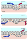
Three models of IFITM protein transmembrane topology. (a) The predicted type III transmembrane topology with endoplasmic reticulum–luminal N and C termini. In support of this model, several flow cytometry studies have localized IFITM1 or IFITM3 N termini to the cellular exterior (7, 45, 58, 61). (b) An intramembrane topology was proposed based on the findings of posttranslational modifications. Ubiquitylation and phosphorylation of NTD amino acids support cytosolic localization of the N terminus (49, 59). Palmitoylation of a C-terminal cysteine in the short TM2/IM2 region of mouse Ifitm1 is consistent with a cytosolic orientation of the C terminus (60). (c) More recent work has shown that murine Ifitm3 can adopt at least two different topologies. The predominant topology, however, has an intracellular N terminus and extracellular C terminus (61). Abbreviations: CIL, conserved intracellular loop; CTD, C-terminal domain; IMD, intramembrane domain (previously denoted as IM1 or TM1); NTD, N-terminal domain; TMD, transmembrane domain (previously denoted as IM2 or TM2).
Figure 4
Figure 4
IFITM protein localization depends on the particular IFITM protein, cell type, and expression level. (a) A549 cells were transduced with an N-terminally Myc-tagged IFITM3 construct. Myc-IFITM3 (red) colocalizes with endogenous CD63 (green) in cytoplasmic puncta. Not all IFITM3-positive vesicles are CD63 positive, and not all CD63-positive vesicles are IFITM3 positive. In cells expressing high levels of the protein (marked by asterisks), IFITM3 is visible at the plasma membrane. (b) A549 cells were transduced with Myc-tagged IFITM1 and stained for Myc (red) and endogenous LAMP2 (green). IFITM1 expression caused marked enlargement of the LAMP2-positive vesicles. (c) Laser scanning confocal microscopy of influenza A virus (IAV)-infected mouse lung parenchyma revealed that alveolar type II pneumocytes express Ifitm3 (red) on lysosome-related organelles [lamellar bodies marked by DC-LAMP (green)]. Punctate cytoplasmic distribution of Ifitm3 also is seen in adjacent type I pneumocytes. (d) Ifitm3 is localized to the cilia and apical plasma membrane of ciliated columnar epithelial cells of the upper airways. The left panel shows a phase-contrast image of two ciliated cells (cilia indicated by arrows) from a bronchus of an IAV-infected mouse. In the right panel, staining for Ifitm3 (red) and lysosomal antigen MAC-3 (green) has been superimposed onto the image. (e) Electron micrograph (5,000× magnification) of a Vero E6 cell overexpressing human IFITM1. The enlarged vesicles (marked by an asterisk) are mostly empty. (f) Electron micrograph (5,000× magnification) of a Vero E6 cell overexpressing human IFITM3. The enlarged vesicles (marked by asterisks) are packed with round membranous structures consistent with intraluminal vesicles.

Similar articles

Cited by

References

    1. Diamond MS, Farzan M. The broad-spectrum antiviral functions of IFIT and IFITM proteins. Nat Rev Immunol. 2013;13:46–57. - PMC - PubMed
    1. Duggal NK, Emerman M. Evolutionary conflicts between viruses and restriction factors shape immunity. Nat Rev Immunol. 2012;12:687–95. - PMC - PubMed
    1. Bieniasz PD. Intrinsic immunity: a front-line defense against viral attack. Nat Immunol. 2004;5:1109–15. - PubMed
    1. Schoggins JW, Wilson SJ, Panis M, Murphy MY, Jones CT, et al. A diverse range of gene products are effectors of the type I interferon antiviral response. Nature. 2011;472:481–85. - PMC - PubMed
    1. Friedman RL, Manly SP, McMahon M, Kerr IM, Stark GR. Transcriptional and posttranscriptional regulation of interferon-induced gene expression in human cells. Cell. 1984;38:745–55. - PubMed