. 2014 Nov 1;1(1):37-45.
doi: 10.1016/j.ebiom.2014.10.022.

Vaccine-induced Human Antibodies Specific for the Third Variable Region of HIV-1 gp120 Impose Immune Pressure on Infecting Viruses

Affiliations

Vaccine-induced Human Antibodies Specific for the Third Variable Region of HIV-1 gp120 Impose Immune Pressure on Infecting Viruses

Susan Zolla-Pazner et al. EBioMedicine. .

Abstract

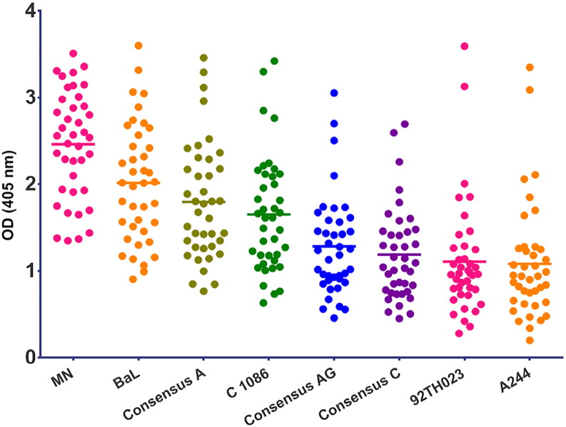
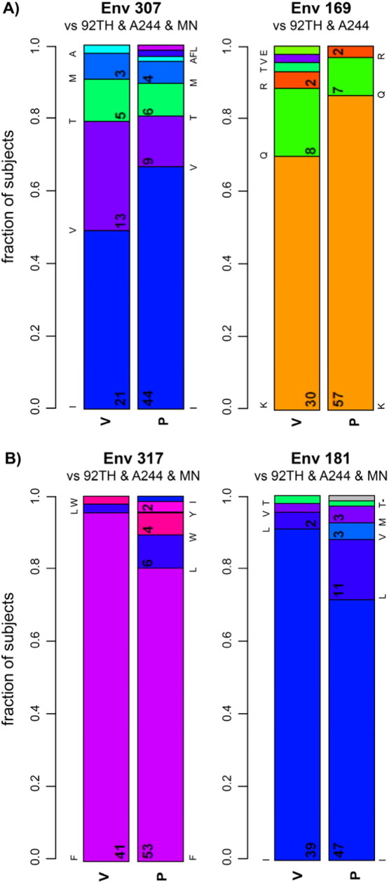
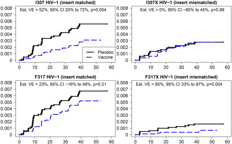
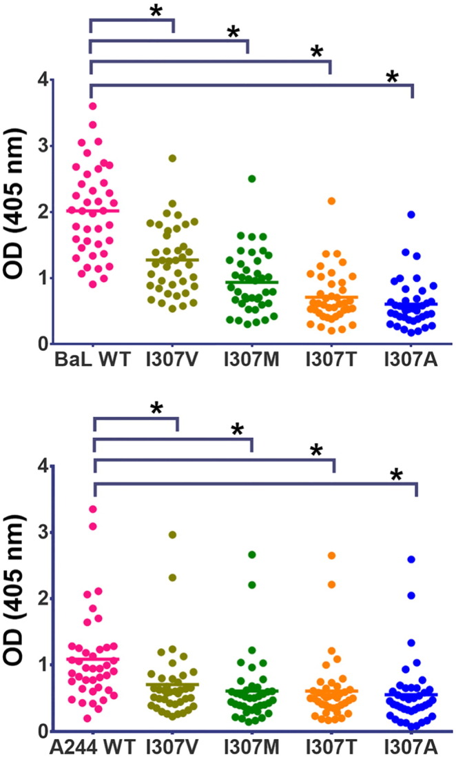
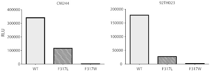
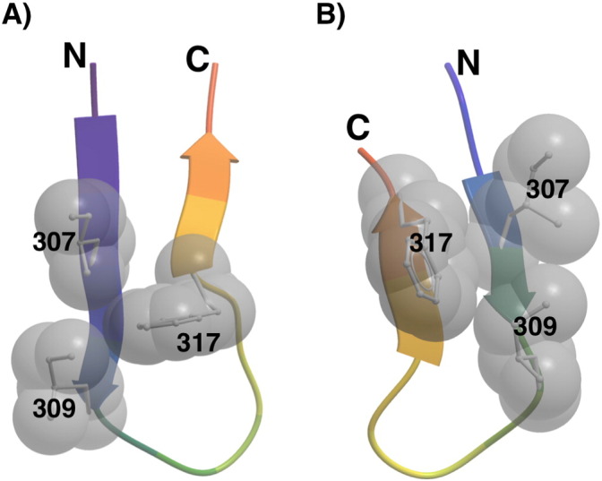
To evaluate the role of V3-specific IgG antibodies (Abs) in the RV144 clinical HIV vaccine trial, which reduced HIV-1 infection by 31.2%, the anti-V3 Ab response was assessed. Vaccinees' V3 Abs were highly cross-reactive with cyclic V3 peptides (cV3s) from diverse virus subtypes. Sieve analysis of CRF01_AE breakthrough viruses from 43 vaccine- and 66 placebo-recipients demonstrated an estimated vaccine efficacy of 85% against viruses with amino acids mismatching the vaccine at V3 site 317 (p=0.004) and 52% against viruses matching the vaccine at V3 site 307 (p=0.004). This analysis was supported by data showing vaccinees' plasma Abs were less reactive with I307 replaced with residues found more often in vaccinees' breakthrough viruses. Simultaneously, viruses with mutations at F317 were less infectious, possibly due to the contribution of F317 to optimal formation of the V3 hydrophobic core. These data suggest that RV144-induced V3-specific Abs imposed immune pressure on infecting viruses and inform efforts to design an HIV vaccine.

Keywords: HIV; antibody; clinical trial; vaccine.


Figures

Fig. 1
Fig. 1
ELISA reactivity with cyclic V3 peptides of RV144 vaccinees' plasma (n = 40) drawn at week 26 (two weeks after the last immunization). The y-axis shows optical density readings at 405 nm for each plasma specimen against a given cyclic V3 peptide (identified on the x-axis). Plasma from placebo recipients were non-reactive and are not shown. All the cyclic V3 peptides were tested in two to three experiments, with two replicates in each experiment; a representative experiment is shown. As shown in Table S1, each group was significantly different from one another (Holm adjusted p-values less than 0.05) except for Conc C vs. A244 (p = 0.086), Conc C vs. 92TH023 (p = 0.16) and 92TH023 vs. A244 (p = 0.16).
Fig. 2
Fig. 2
Fraction of viruses from vaccinees and placebo recipients with designated residues at V3 position 307 and V2 position 169 (A), and with designated residues at V3 position 317 and V2 position 181 (B). The vaccine immunogen amino acid is represented by the bottom-most bar in each plot. For all positions except Env 169, the amino acid was the same in all three immunogen sequences; at Env 169 the MN immunogen AA (methionine) was not found in any subject's breakthrough sequence so it is not depicted.
Fig. 3
Fig. 3
Estimated cumulative incidences of HIV-1 infection in placebo and vaccine recipients. The probability of acquiring HIV infection (y-axis) is shown for vaccine and placebo recipients whose viruses contained residues at V3 positions 307 (upper panels) or 317 (lower panels) which were either matched (I307 or F317; left panels) or mismatched (I307X or F317X; right panels) to the residues at these positions in the vaccine. The x-axis shows months since entry into the study.
Fig. 4
Fig. 4
Reactivity of plasma from RV144 vaccinees (week 26) with wild type cyclic V3 peptides from clade B (strain BaL: upper panel) and clade AE (strain A244: lower panel) compared to variants with amino acid substitutions at position 307. All cyclic V3 peptides were tested in two to three experiments, with two replicates in each experiment. Asterisks denote p < 0.0001 by paired t-test.
Fig. 5
Fig. 5
Infectivity of wild type (WT) CM244 and 92TH023 pseudoviruses and pseudoviruses with an F317L mutation or an F317W mutation.
Fig. 6
Fig. 6
Structure of the V3 hydrophobic core. The side chains of V3 residues I307, I309, and F317 often pack against each other to form a hydrophobic core and are spatially located at either side of the hairpin turn in the V3 crown as represented by the V3 structures in complex with V3-specific mAb 447-52D (A) (Killikelly et al., 2013) or mAb 2557 (B) (Jiang et al., 2010). The V3 hairpin is drawn as a ribbon, and the side chains (toward the reader) of I307, I309 and F317 as sticks and CPK spheres.

References

    1. Abagyan R., Batalov S. Homology modeling with internal coordinate mechanics: deformation zone mapping and improvements of models via conformational search. Proteins. 1997;(Suppl. 1):29–37. - PubMed
    1. Almond D., Kimura T. Structural conservation predominates over sequence variability in the crown of HIV-1'S V3 loop. AIDS Res. Hum. Retroviruses. 2010;26(6):717–723. - PMC - PubMed
    1. Andrabi R., Williams C. Cross-neutralizing activity of human anti-V3 monoclonal antibodies derived from non-B clade HIV-1 infected individuals. Virology. 2013;439(2):81–88. - PMC - PubMed
    1. Andrus L., Prince A.M. Passive immunization with a human immunodeficiency virus type-1 neutralizing monoclonal antibody in Hu-PBL-SCID mice: isolation of a neutralization escape variant. J. Infect. Dis. 1998;177(4):889–897. - PubMed
    1. Bartesaghi A., Merk A. Prefusion structure of trimeric HIV-1 envelope glycoprotein determined by cryo-electron microscopy. Nat. Struct. Mol. Biol. 2013;20(12):1352–1357. - PMC - PubMed

LinkOut - more resources