

Circulating E3 ligases are novel and sensitive biomarkers for diagnosis of acute myocardial infarction
- PMID: 25599194
- PMCID: PMC4557400
- DOI: 10.1042/CS20140663
Circulating E3 ligases are novel and sensitive biomarkers for diagnosis of acute myocardial infarction
Abstract
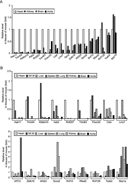
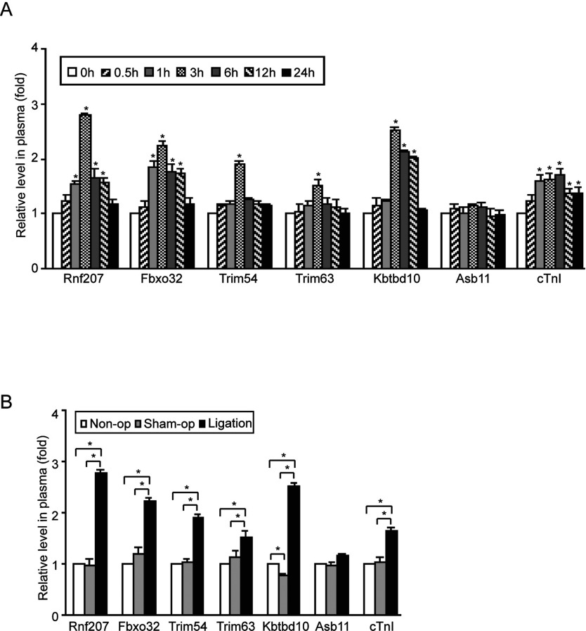
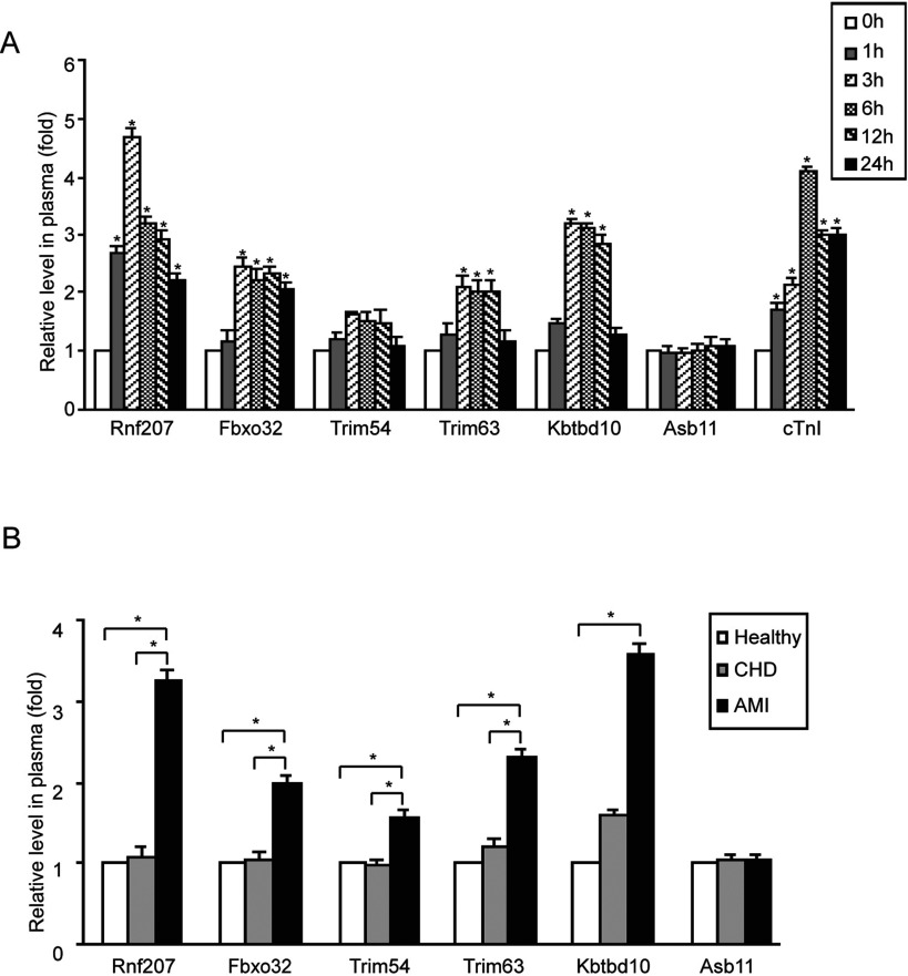
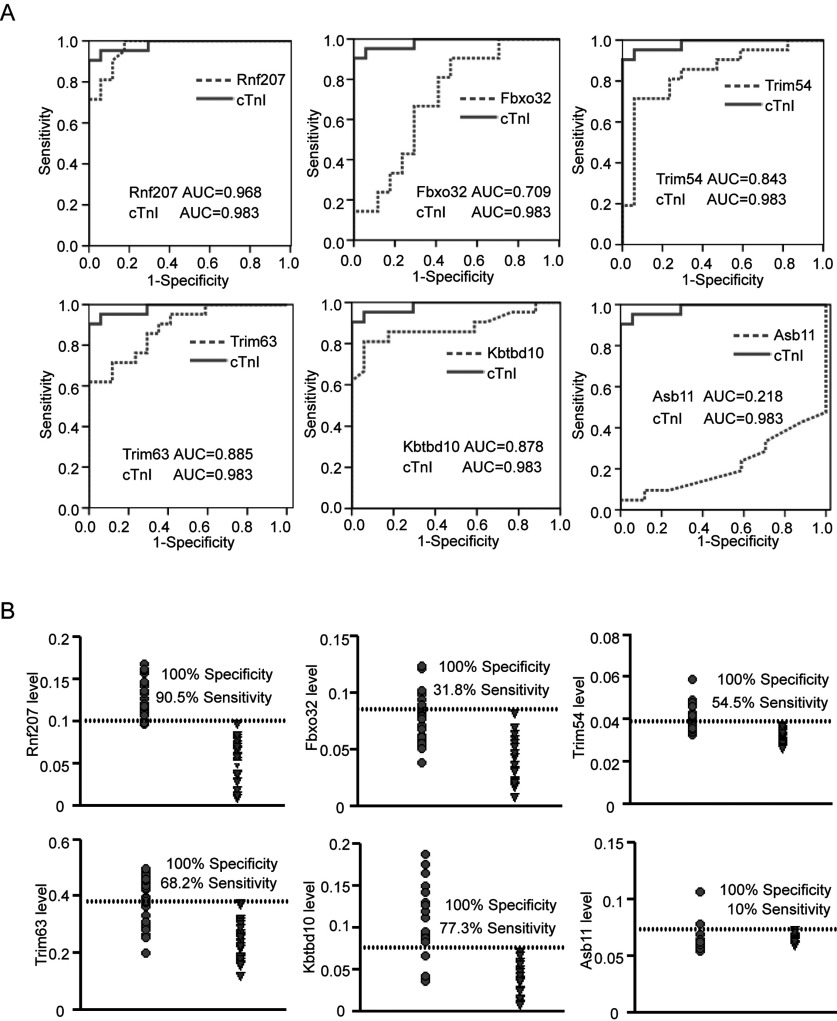
Ubiquitin ligase (E3) is a decisive element of the ubiquitin-proteasome system (UPS), which is the main pathway for intracellular protein turnover. Recently, circulating E3 ligases have been increasingly considered as cancer biomarkers. In the present study, we aimed to determine if cardiac-specific E3 ligases in circulation can serve as novel predictors for early diagnosis of acute myocardial infarction (AMI). By screening and verifying their tissue expression patterns with microarray and real-time PCR analysis, six of 261 E3 ligases, including cardiac-specific Rnf207 and cardiac- and muscle-enriched Fbxo32/atrogin-1, Trim54/MuRF3, Trim63/MuRF1, Kbtbd10/KLHL41, Asb11 and Asb2 in mouse heart, were selected for the present study. In the AMI rats, the levels of five E3 ligases including Rnf207, Fbxo32, Trim54, Trim63 and Kbtbd10 in the plasma were significantly increased compared with control animals. Especially, the plasma levels of Rnf207 was markedly increased at 1 h, peaked at 3 h and decreased at 6-24 h after ligation. Further evaluation of E3 ligases in AMI patients confirmed that plasma Rnf207 level increased significantly compared with that in healthy people and patients without AMI, and showed a similar time course to that in AMI rats. Simultaneously, plasma level of cardiac troponin I (cTnI) was measured by ELISA assays. Finally, receiver operating characteristic (ROC) curve analysis indicated that Rnf207 showed a similar sensitivity and specificity to the classic biomarker troponin I for diagnosis of AMI. Increased cardiac-specific E3 ligase Rnf207 in plasma may be a novel and sensitive biomarkers for AMI in humans.
Figures
References
-
- Go A.S., Mozaffarian D., Roger V.L., Benjamin E.J., Berry J.D., Borden W.B., Bravata D.M., Dai S., Ford E.S., Fox C.S., et al. Executive summary: heart disease and stroke statistics–2013 update: a report from the American Heart Association. Circulation. 2013;127:143–152. doi: 10.1161/CIR.0b013e318282ab8f. - DOI - PubMed
-
- Alpert J.S., Thygesen K., Antman E., Bassand J.P. Myocardial infarction redefined—a consensus document of The Joint European Society of Cardiology/American College of Cardiology Committee for the redefinition of myocardial infarction. J. Am. Coll. Cardiol. 2000;36:959–969. doi: 10.1016/S0735-1097(00)00804-4. - DOI - PubMed
Publication types
MeSH terms
Substances
LinkOut - more resources
Full Text Sources
Medical
Molecular Biology Databases
Research Materials