Review
. 2015 Jan;261(1):46-55.
doi: 10.1097/SLA.0000000000000622.

The status of contemporary image-guided modalities in oncologic surgery

Affiliations
Review

The status of contemporary image-guided modalities in oncologic surgery

Eben L Rosenthal et al. Ann Surg. 2015 Jan.

Abstract

Objective: To review the current trends in optical imaging to guide oncologic surgery.

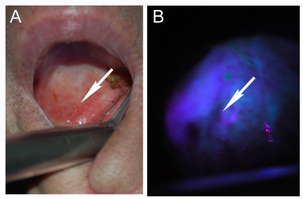
Background: Surgical resection remains the cornerstone of therapy for patients with early stage solid malignancies and more than half of all patients with cancer undergo surgery each year. The technical ability of the surgeon to obtain clear surgical margins at the initial resection remains crucial to improve overall survival and long-term morbidity. Current resection techniques are largely based on subjective and subtle changes associated with tissue distortion by invasive cancer. As a result, positive surgical margins occur in a significant portion of tumor resections, which is directly correlated with a poor outcome.

Methods: A comprehensive review of studies evaluating optical imaging techniques is performed.

Results: A variety of cancer imaging techniques have been adapted or developed for intraoperative surgical guidance that have been shown to improve functional and oncologic outcomes in randomized clinical trials. There are also a large number of novel, cancer-specific contrast agents that are in early stage clinical trials and preclinical development that demonstrate significant promise to improve real-time detection of subclinical cancer in the operative setting.

Conclusions: There has been an explosion of intraoperative imaging techniques that will become more widespread in the next decade.


Figures

Figure 1
Figure 1. Sampling error in oncologic surgery
An unresected tongue cancer (upper left pane) requires circumferential mucosal margin assessment that would require almost 8 cm of healthy tissue to be sampled in order to confirm a negative margin (upper right). Once resected (lower left) the wound bed requires almost 40 samples of tissue within the three dimensional cavity (lower right). Geographic mapping of the sample location is complicated by the size and mobility of the defect.
Figure 2
Figure 2. Tissue interaction with light
(A) Light as it enters a medium such as tissue will be reflected and succumbs to scattering that limits penetration. Light can also be absorbed by molecules within the tissue (chromophores) or excite endogenous or exogenously administered molecules to emit light at a different wavelength. (B) Penetration of light through tissue is dependent on the absorptive properties of the tissue at various wavelengths. Near infrared (NIR) light has the best penetration through soft-tissue.
Figure 3
Figure 3. Autofluorescence has been adopted for screening and identification of margins in oral cavity cancer
Blue/violet light is applied to the mucosa surfaces (A) and areas of decreased autofluorescence correlate with inflamed or neoplastic changes (B).
Figure 4
Figure 4. Multi-modality imaging techniques adopted in the surgical suite
(a) Preoperative contrast-enhanced T1-weighted MR images show patchy/faint CE and (g) hyperintensity on FLAIR sequences. (b) The intratumoral area outside the region of maximum positron emission tomography (PET) tracer uptake verified by the intraoperative navigation system (c) appeared as whitish glioma tissue under the surgical microscope, (d) with no detectable PpIX fluorescence. (e) The corresponding histopathology reveals low-grade glioma tissue according to the WHO criteria in the H&E stain (f) with a low proliferation rate (MIB-1: <10%). (h) In contrast, the intratumoral area inside the region of maximum PET tracer uptake (i) showed similar glioma tissue appearance in the microscopic view, (j) but revealed strong PpIX fluorescence under violet-blue excitation light. (k) The corresponding histopathology reveals high-grade glioma tissue in accordance with an anaplastic focus according to the WHO criteria in the H&E stain (l) with a high proliferation rate (MIB-1: 32%). The final histopathological diagnosis revealed a focally anaplastic astrocytoma (WHO grade III) and the patient was treated with radiochemotherapy. The width of each histopathological image (e, f, k, l) was 300 micrometers (μm). Image and legend information was taken from Widhalm et al, PLoS ONE 2013.
Figure 5
Figure 5. Small fragments of tumor have tissue penetration of greater than 5 mm
Layers of skin were used to measure the penetration of a small tumor fragment (18 mm3). A mouse bearing a head and neck tumor was injected with cetuximab conjugated to a NIR dye (emission 705 nm) and then the skin and tumor fragments harvested and imaged by stereomicroscopy as above ex vivo. Three layers of skin alone (A, D) had minimal fluorescence. Tumor was clearly visualized alone (B, E) or with layers of overlying skin (C, F). Notice significant scattering associated with overlying tissue.
Figure 6
Figure 6. Intraoperative imaging of tumors using ICG
A) Intraoperative imaging of tongue cancer under white light using the OH5 surgical microscope (Leica). B) After systemic injection of 7 mg of indocyanine green the Leica FL800 microscope can differentiate tumor border. Untargeted indocyanine green accumulates within the tumor based on enhanced permeability and retention effect of cancer and the high blood flow in tumors, but is otherwise non-specific.

References

    1. Woolgar JA, Triantafyllou A. A histopathological appraisal of surgical margins in oral and oropharyngeal cancer resection specimens. Oral Oncol. 2005;41:1034–43. - PubMed
    1. McMahon J, O’Brien CJ, Pathak I, et al. Influence of condition of surgical margins on local recurrence and disease-specific survival in oral and oropharyngeal cancer. Br J Oral Maxillofac Surg. 2003;41:224–231. - PubMed
    1. Ravasz LA, Slootweg PJ, Hordijk GJ, et al. The status of the resection margin as a prognostic factor in the treatment of head and neck carcinoma. J Craniomaxillofac Surg. 1991;19:314–318. - PubMed
    1. Atkins J, Al Mushawah F, Appleton CM, et al. Positive margin rates following breast-conserving surgery for stage I-III breast cancer: palpable versus nonpalpable tumors. J Surg Res. 2012;177:109–115. - PMC - PubMed
    1. Iczkowski KA, Lucia MS. Frequency of positive surgical margin at prostatectomy and its effect on patient outcome. Prostate Cancer. 2011;2011:673021. - PMC - PubMed

Publication types