. 2015 Feb;17(2):113-22.
doi: 10.1038/ncb3091. Epub 2015 Jan 19.

Lineage specificity of primary cilia in the mouse embryo

Affiliations

Lineage specificity of primary cilia in the mouse embryo

Fiona K Bangs et al. Nat Cell Biol. 2015 Feb.

Abstract

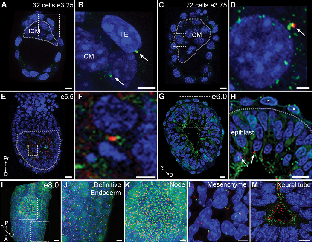
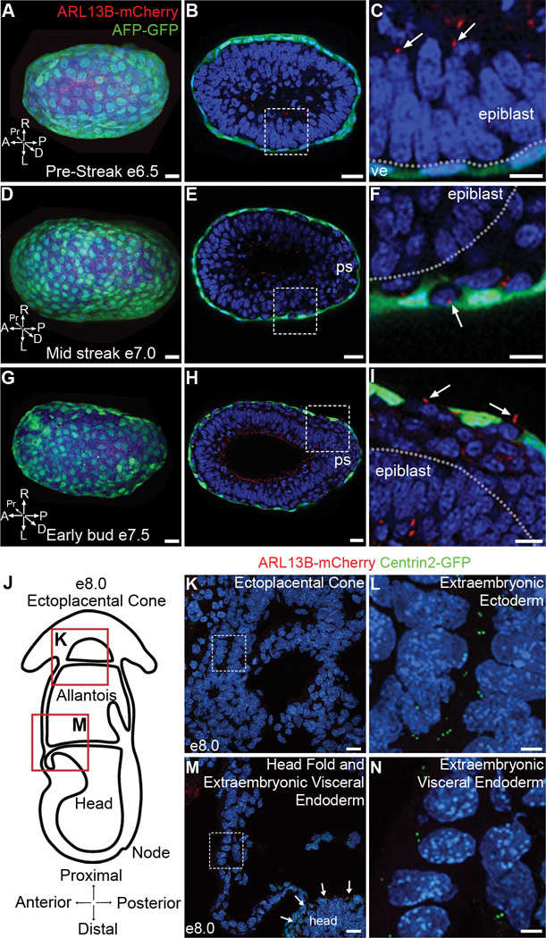
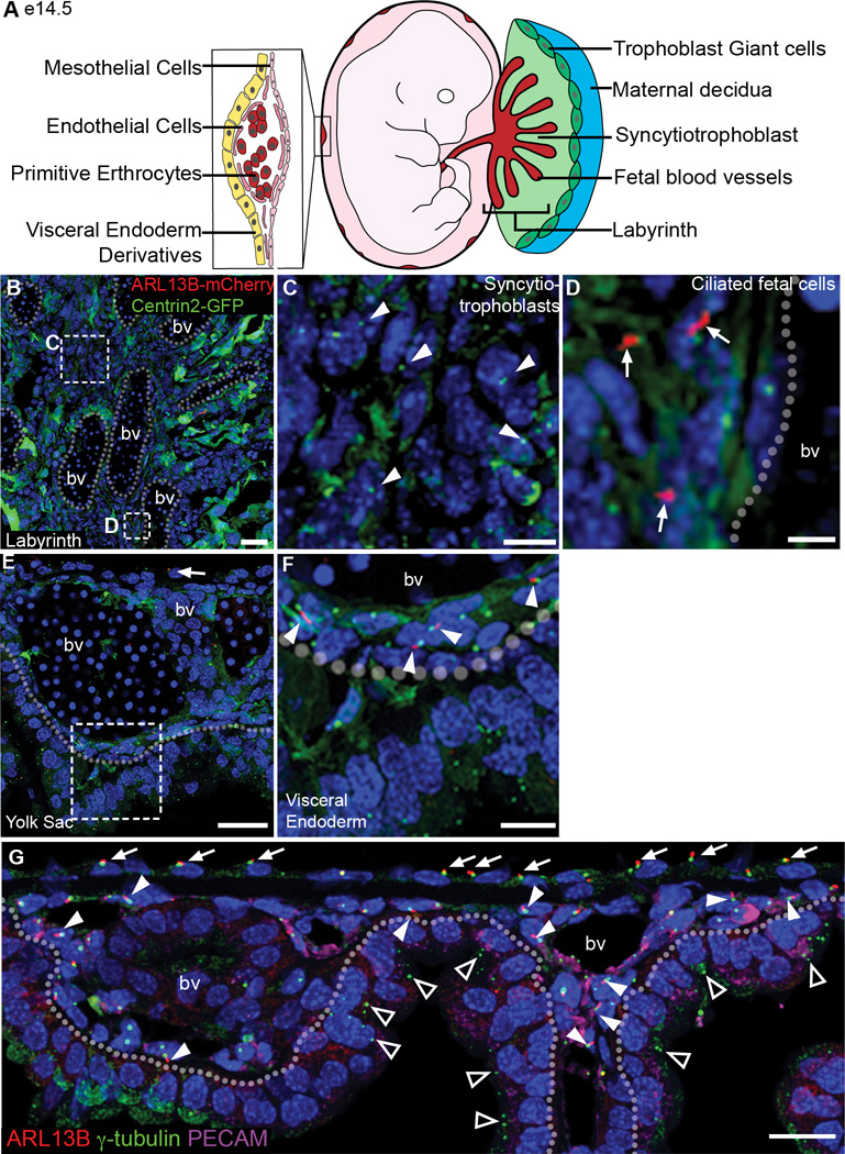
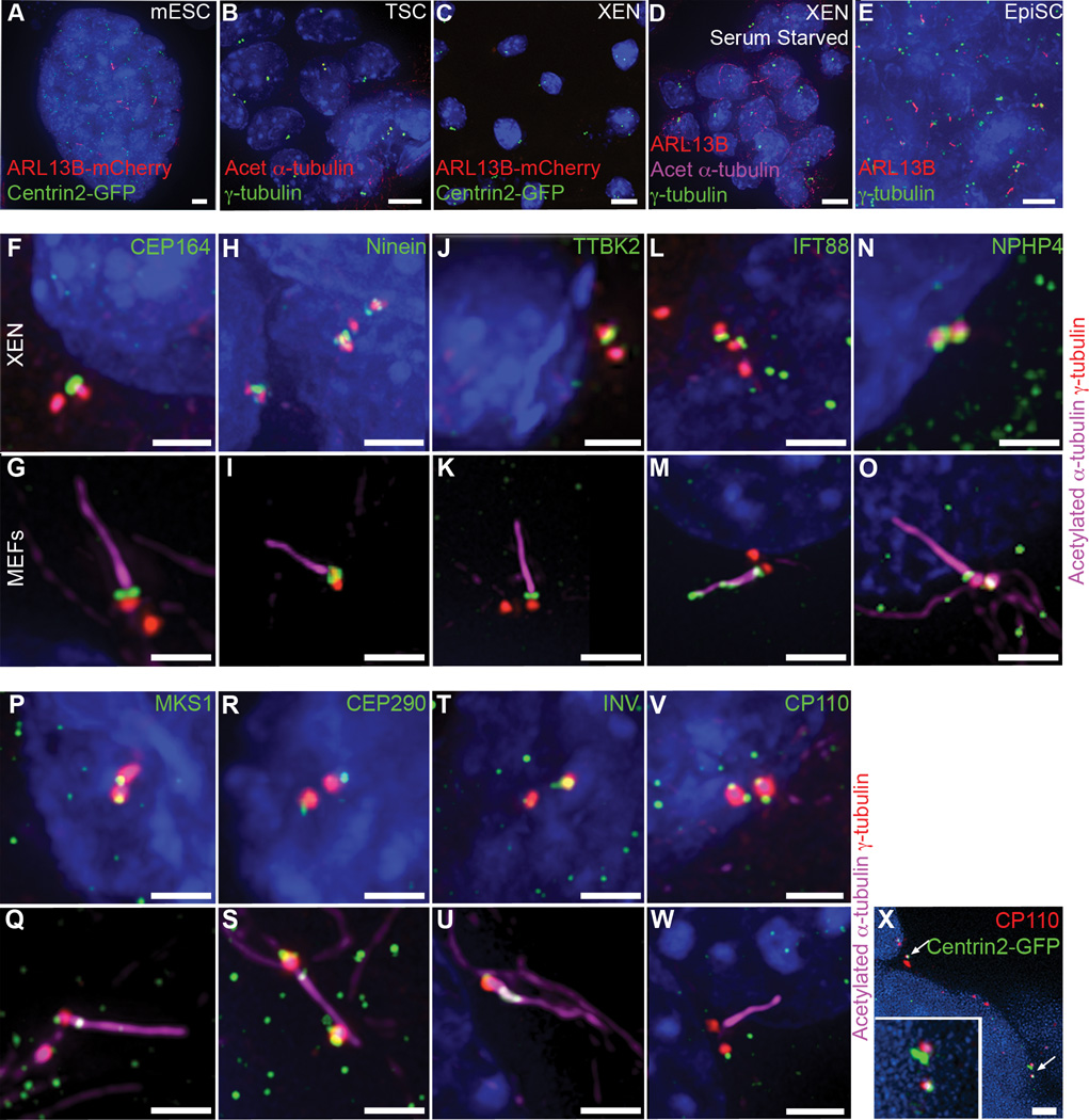
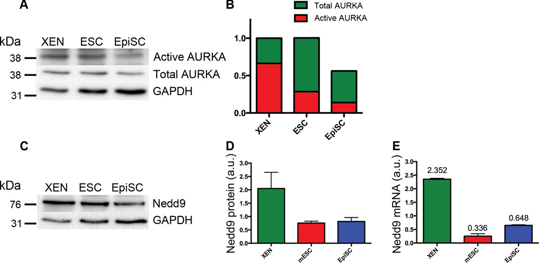
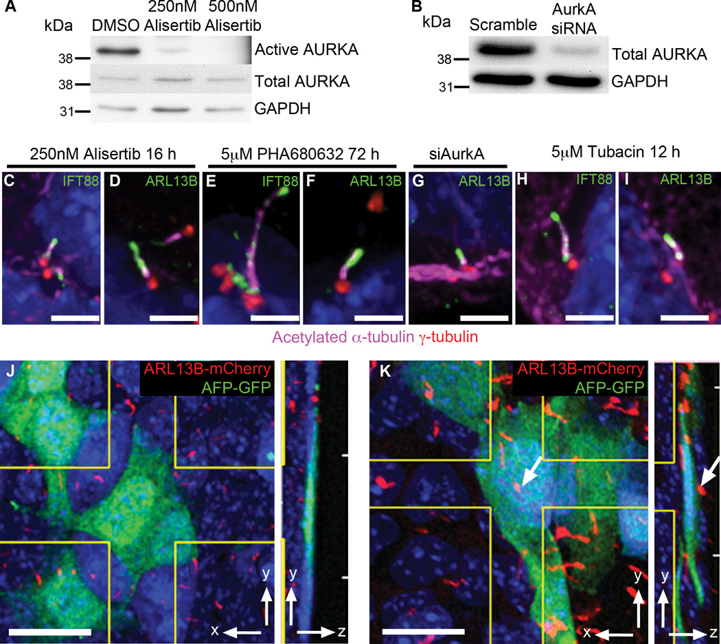
Primary cilia are required for vertebrate cells to respond to specific intercellular signals. Here we define when and where primary cilia appear in the mouse embryo using a transgenic line that expresses ARL13B-mCherry in cilia and Centrin 2-GFP in centrosomes. Primary cilia first appear on cells of the epiblast at E6.0 and are subsequently present on all derivatives of the epiblast. In contrast, extraembryonic cells of the visceral endoderm and trophectoderm lineages have centrosomes but no cilia. Stem cell lines derived from embryonic lineages recapitulate the in vivo pattern: epiblast stem cells are ciliated, whereas trophoblast stem cells and extraembryonic endoderm (XEN) stem cells lack cilia. Basal bodies in XEN cells are mature and can form cilia when the AURKA-HDAC6 cilium disassembly pathway is inhibited. The lineage-dependent distribution of cilia is stable throughout much of gestation, defining which cells in the placenta and yolk sac are able to respond to Hedgehog ligands.


Figures

Figure 1
Figure 1. Primary cilia arise in the post-implantation epiblast
Embryos homozygous for the ARL13B-mCherry (red) and Centrin2-GFP (green) transgenes reveal the distribution of centrosomes and primary cilia in the early mouse embryo. (A–D) Preimplantation embryos. (A) Optical section of 32 cell early blastocyst (e3.25); dotted line surrounds ICM. (B) Expanded view of the dashed box in (A) shows that Centrin2-GFP positive centrioles are present on both trophectoderm cells (TE) and inner cell mass cells (ICM), arrows. No primary cilia are observed (Representative image selected from 4 embryos). (C) Optical section of a 72 cell blastocyst (e3.75); dotted line surrounds ICM. No cilia were detected on trophectoderm cells (0 cilia/255 cells from 9 embryos). (D) Expanded view of dashed box shown in (C) shows a rare example (2/107 cells scored from 9 embryos) of ARL13B-mCherry (arrow) expression adjacent to a Centrin2-GFP+ centriole of an ICM cell; these puncta did not have the morphology of cilia nor did they express acetylated α-tubulin. (E–H) Early postimplantation embryos. (E) Longitudinal optical section of e5.5 cavitating embryo; dotted line surrounds the epiblast. (F) Expanded view of the box in (E) shows a rare cilium on an epiblast cell (3/654 cells from 10 embryos). (G) Transverse optical section on an e6.0 embryo, with cilia exclusively on epiblast cells (388/1039 cells from 6 embryos). (H) Expanded view of dashed box in (G) shows cilia (arrows) on epiblast cells; no cilia are present on Centrin2-GFP labeled centrioles in visceral endoderm cells (asterisks; 0/837 cells from 6 embryos). Dotted line denotes boundary between epiblast (below) and visceral endoderm (above). (I) Primary cilia are detected on nearly all cells of the e8.0 embryo. (J, K) Expanded views of dashed boxes in (I) show the cilia in the definitive endoderm (J) and node (K). (L, M) Sections of e10.5 embryos. Cilia are detected in nearly all cells in the limb mesenchyme (L). Neural progenitors extend cilia into the lumen of the neural tube (M). Nuclei are marked with DAPI (blue). Scale bars: (A, B): 10 µm; (C): 8 µm; (D): 2 µm (E): 20 µm; (F): 5 µm; (G, H): 20 µm; (I) 30 µm; (J, K): 10 µm; (L, M) 5 µm. P = posterior, A = anterior, Pr = proximal, D = distal.
Figure 2
Figure 2. Primary cilia are restricted to embryonic lineages in late gastrulation stage embryos
(A–C) Primary cilia labeled with ARL13B-mCherry (red) are present on epiblast cells but are absent from cells of the visceral endoderm marked with AFP-GFP (green). (A) Distal view of pre-streak stage embryo (e6.5; Representative image selected from 4 embryos) expressing AFP-GFP and ARL13B-mCherry. (B) Transverse optical section of embryo in (A). (C) Magnification of dashed box in (B) shows presence of primary cilia labeled with ARL13B-mCherry (red) on epiblast cells, (arrow), but absent from visceral endoderm cells (ve), labeled with AFP-GFP (green). Dotted line denotes boundary between epiblast (above) and visceral endoderm (below). (D–I) As the definitive endoderm intercalates with the visceral endoderm (green), cilia (red) are present on the definitive, but not the visceral, endoderm. (D) Distal view of mid-streak stage embryo (e7.0; Representative image selected from 3 embryos) expressing AFP-GFP and ARL13B-mCherry. (E) Transverse optical section of embryo in (D), primitive streak (ps) is to the right. (F) Magnification of dashed box in (E) shows that primary cilia are present on definitive endoderm cells, (arrow), but absent from surrounding visceral endoderm cells, labeled with AFP-GFP (green). Dotted line denotes boundary between epiblast and definitive endoderm. (G) Distal view of early bud stage embryo (e7.5) expressing AFP-GFP and ARL13B-mCherry (Representative image selected from 3 embryos). (H) Transverse optical section of embryo in (G), primitive streak (ps) is to the right. (I) Expanded view of box in (H) shows primary cilia on definitive endoderm cells (arrows) that have intercalated between AFP-GFP+ visceral endoderm cells (green). Dotted line denotes boundary between epiblast (below) and definitive endoderm (above). P = posterior, A = anterior, Pr = proximal, D = distal, L = left, R = right. (J) Schematic of e8.0 embryo in cross section illustrates the locations of images shown in (K) and (M). (K) Cells of the ectoplacental cone (derivatives of trophoblast lineage) expressing ARL13B-mCherry and Centrin2-GFP. (L) Magnification of dashed box in (K) shows Centrin2-GFP labeled centrioles with no primary cilium on extraembryonic ectoderm cells. (M) Cells of the extraembryonic visceral endoderm expressing ARL13B-mCherry and Centrin2-GFP are not ciliated, although epiblast cells in the headfolds are ciliated, arrows. (N) Magnification of dashed box (M) shows Centrin2-GFP labeled centrioles but no primary cilia on extraembryonic visceral endoderm cells. Nuclei are marked with DAPI (blue). Scale bars: (A–F) 30 µm; (G–I) 40 µm; (K, M) 20 µm; (L, N) 5 µm.
Figure 3
Figure 3. Extraembryonic lineages of the placenta and yolk sac lack cilia at e14.5
(A) Schematics of the placenta and yolk sac: trophoblast giant cells (dark green), spongiotrophoblast (light green), labyrinth (red), and magnified view of the yolk sac, illustrating a blood vessel surrounded by endothelial cells (dark pink) and the positions of mesothelial cells (light pink) and extraembryonic visceral endoderm derivatives (yellow). (B) Section of e14.5 labyrinth layer from an ARL13B-mCherry Centrin2-GFP placenta; dotted lines outline fetal blood vessels (bv). (C) Magnified view of box in (B), non-ciliated trophoblast-derived syncytiotrophoblast cells between fetal blood vessels lack cilia; arrowheads indicate Centrin2-GFP+ centrioles. (D) Magnified view of box in (B), showing cilia on mesoderm derived cells surrounding fetal blood vessels (arrows). (E) Section of e14.5 yolk sac expressing ARL13B-mCherry and Centrin2-GFP; dashed line demarcates the boundary between mesoderm-derived cells (mesothelial and endothelial cells, above) and extraembryonic visceral endoderm-derived cells (below). Arrow indicates ciliated mesothelial cell. (F) Magnified view of box in (E) shows ciliated endothelial cells, (arrows) while extraembryonic visceral endoderm cells, below dashed line, are not ciliated. (F) Section of e14.5 yolk sac; dotted line demarcates the boundary between mesoderm-derived cells (mesothelial and endothelial cells, above) and extraembryonic visceral endoderm-derived cells (below). Antibody staining for γ-tubulin, labels centrosomes (green), and ARL13B, labels cilia (red), are present on mesothelial cells (arrows), and PECAM-expressing (magenta) endothelial cells (arrow heads). Extraembryonic visceral endoderm-derived cells have centrosomes (triangles) but no cilia (221/386 mesothelial cells; 95/292 endothelial cells; 0/1032 extraembryonic visceral endoderm-derived cells from 2 embryos). bv= blood vessel. Nuclei are marked with DAPI (blue). Scale bars: (B) 30 µm; (C,D) 10 µm; (E) 30 µm; (F) 10 µm; (G) 20 µm.
Figure 4
Figure 4. Embryo-derived stem cells recapitulate the cilia status of embryonic lineages
(A–E) Presence of primary cilia on embryo-derived stem cells. (A) 18% of asynchronously dividing mESCs derived from ARL13B-mCherry (red) Centrin2-GFP (green) transgenic embryos grown in 2i medium are ciliated. (B) Antibody staining for γ-tubulin (green) and acetylated α-tubulin (red) shows TS cells lack primary cilia. (C) No cilia are detected on XEN cells derived from ARL13B-mCherry Centrin2-GFP transgenic embryos. (D) Antibody staining for ARL13B (red) and acetylated α-tubulin (magenta) shows that serum starved XEN cells lack cilia (0/267 cells from 2 independent experiments). (E) Antibody staining of EpiSCs for γ-tubulin (green) and ARL13B (red) shows that almost all EpiSCs are ciliated. (F–U) XEN cells have mature basal bodies. Centrioles marked with γ-tubulin (red) are associated with the distal appendage marker Cep164 (green) (F) and subdistal appendage marker ninein (green)(H). Positive regulators of ciliogenesis TTBK2 (green) (J) and IFT88 (green) (L) as well as transition zone proteins (green) NPHP4 (N), MKS1 (P), CEP290 (R) and Inversin (T) are also present at the mother centriole in XEN cells. (G, I, K, M, O, Q, S, U) Localization of basal body proteins in ciliated mouse embryonic fibroblasts (MEFs) is the same as in XEN cells, although IFT88 is also present in the axoneme in MEFs marked with acetylated α-tubulin (magenta). The negative regulator of ciliogenesis CP110 (green) is present on both centrioles in all XEN cells (V) but is removed from the mother centriole upon cilia assembly in MEFs (W). CP110 (red) is also present on both centrioles marked with Centrin2-GFP (green) in cells of the embryonic visceral endoderm (X, arrows). Nuclei are marked with DAPI (blue). Scale bars: (A–E) 7 µm; (F–W) 2 µm; (X) 3 µm.
Figure 5
Figure 5. Components of the cilia disassembly pathway are highly expressed in XEN cells
(A) Western blot analysis of activated and total AURKA. (B) Quantification of the western blots for total AURKA (green) and activated AURKA (red), (n=3 independent experiments, normalized to XEN). (C) Western blot analysis of NEDD9 protein levels. (D) Quantification of Western blot analysis (C) shows that NEDD9 protein levels are 2.64 ± 1.15 fold higher in XEN cells compared to EpiSCs (n=3 independent experiments). Error bars indicate standard deviation. (E) Nedd9 mRNA levels are higher in XEN cells than in EpiSCs by qPCR (values indicate mean mRNA expression levels, n=3 independent experiments). Error bars indicate standard deviation.
Figure 6
Figure 6. XEN and VE cells form cilia when the cilia disassembly pathway is inhibited
(A) Western blot shows that treatment for 16 h with 250 nM Alisertib reduces levels of phosphorylated AurkA by 32.5-fold compared to DMSO controls. Phosphorylated AurkA was not detected in cells treated with 500 nM Alisertib, whereas total AurkA levels are unaffected (n=2 independent experiments). (B) Western blot analysis shows that siRNA knock down of AurkA reduces levels of total AurkA to ~15 of control levels (n=2 independent experiments). (C, D) Treatment of XEN cells with 250 nM AURKA inhibitor Alisertib for 16h causes formation of primary cilia marked with IFT88 (C, green), ARL13B (D, green), and acetylated α-tubulin (magenta) labeled axoneme projecting from a centrosome marked with γ-tubulin (red; 20/328 cells). (E, F) Treatment of XEN cells with 5 µM AurkA inhibitor PHA680632 for 72 h causes formation of primary cilia marked with IFT88 (E, green), ARL13B (F, green) and acetylated tubulin (magenta) projecting from a centrosome marked with γ-tubulin (red; 15/302 cells). (G) siRNA knock down of AurkA causes formation of primary cilia on XEN cells marked with ARL13B (green) and acetylated α-tubulin (magenta) projecting from a centrosome marked with γ-tubulin (red; 7/610 cells from 2 independent experiments). (H, I) Treatment of XEN cells with 5 µM HDAC6 inhibitor tubacin for 12 h causes formation of primary cilia on XEN cells marked with IFT88 (H), ARL13B (I) and acetylated α-tubulin (magenta), projecting from a centrosome marked with γ-tubulin (red; 8/717 cells from 3 independent experiments). (J, K) Culture of e7.5 embryos for 12h in 5 µM tubacin causes cilia formation (red) on visceral endoderm cells (green). (J) Visceral endoderm cells in an e7.5 embryo expressing AFP-GFP (green) are never ciliated in DMSO treated control embryos (0/1263 cells counted across 6 embryos). (K) Primary cilia, labeled with ARL13B-mCherry (red) are observed on AFP-GFP (green) visceral endoderm cells in embryos cultured with 5 µM tubacin for 12 h (30/1313 cells counted across 7 embryos). Arrow indicates a primary cilium present on this AFP-GFP+ cell. Side panels show yz view. Nuclei stained with DAPI (blue). Scale bars = (C–I) 2 µm; (J, K) 20 µm.

Comment in

References

    1. Goetz SC, Anderson KV. The primary cilium: a signalling centre during vertebrate development. Nat Rev Genet. 2010;11:331–344. - PMC - PubMed
    1. Seeley ES, Nachury MV. The perennial organelle: assembly and disassembly of the primary cilium. J. Cell. Sci. 2010;123:511–518. - PMC - PubMed
    1. Wong SY, et al. Primary cilia can both mediate and suppress Hedgehog pathway–dependent tumorigenesis. Nat Med. 2009;15:1055–1061. - PMC - PubMed
    1. Han Y-G, et al. Dual and opposing roles of primary cilia in medulloblastoma development. Nat Med. 2009;15:1062–1065. - PMC - PubMed
    1. Caspary T, Larkins CE, Anderson KV. The Graded Response to Sonic Hedgehog Depends on Cilia Architecture. Developmental Cell. 2007;12:767–778. - PubMed

Publication types

Substances

LinkOut - more resources