Review
. 2015 Apr;95(4):366-76.
doi: 10.1038/labinvest.2014.153. Epub 2015 Jan 19.

Novel genotype-phenotype associations in human cancers enabled by advanced molecular platforms and computational analysis of whole slide images

Affiliations
Review

Novel genotype-phenotype associations in human cancers enabled by advanced molecular platforms and computational analysis of whole slide images

Lee A D Cooper et al. Lab Invest. 2015 Apr.

Abstract

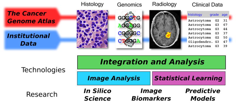
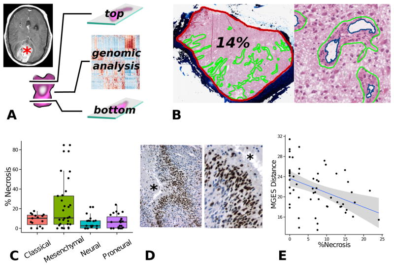
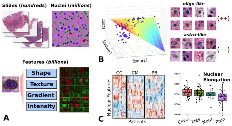
Technological advances in computing, imaging, and genomics have created new opportunities for exploring relationships between histology, molecular events, and clinical outcomes using quantitative methods. Slide scanning devices are now capable of rapidly producing massive digital image archives that capture histological details in high resolution. Commensurate advances in computing and image analysis algorithms enable mining of archives to extract descriptions of histology, ranging from basic human annotations to automatic and precisely quantitative morphometric characterization of hundreds of millions of cells. These imaging capabilities represent a new dimension in tissue-based studies, and when combined with genomic and clinical endpoints, can be used to explore biologic characteristics of the tumor microenvironment and to discover new morphologic biomarkers of genetic alterations and patient outcomes. In this paper, we review developments in quantitative imaging technology and illustrate how image features can be integrated with clinical and genomic data to investigate fundamental problems in cancer. Using motivating examples from the study of glioblastomas (GBMs), we demonstrate how public data from The Cancer Genome Atlas (TCGA) can serve as an open platform to conduct in silico tissue-based studies that integrate existing data resources. We show how these approaches can be used to explore the relation of the tumor microenvironment to genomic alterations and gene expression patterns and to define nuclear morphometric features that are predictive of genetic alterations and clinical outcomes. Challenges, limitations, and emerging opportunities in the area of quantitative imaging and integrative analyses are also discussed.


Conflict of interest statement

CONFLICT OF INTEREST

The authors declare no conflicts of interest.

Figures

Figure 1
Figure 1
Integration of quantitative histology with multifaceted clinical and genomic data. Image analysis algorithms can extract features that describe the histology in digital whole-slide image datasets. This information can be combined with genomic, clinical and radiology data to identify image biomarkers of genetic alterations, to build predictive models of clinical outcomes, and to better understand tumor biology. Public data provided by The Cancer Genome Atlas (TCGA) makes it possible to explore these topics in large cohorts of more than 22 types of cancers. Discoveries made in analysis of public TCGA data can be validated in smaller institutional datasets.
Figure 2
Figure 2
Tumor microenvironment study integrating histology and genomics from TCGA. (A) TCGA specimens are sections from the top and bottom to produce slides, and the middle portion is submitted for genomic analysis. (B) Digitized images from top/bottom sections were annotated to calculate the percentage of necrosis and angiogenesis for each tumor. (C) Tumors from the mesenchymal expression class are significantly enriched with necrosis. (D) As the amount of necrosis increases in non-mesenchymal GBMs, gene expressions patterns shift towards a mesenchymal expression signature.
Figure 3
Figure 3
Quantitative nuclear morphometry. (A) Image analysis algorithms are used to delineate nuclei in whole-slide images. A set of features is calculated to describe the appearance of each nucleus. This system is capable of processing thousands of slides and hundreds of millions of nuclei. (B) We developed a model-based system to score nuclei based on oligodendroglial differentiation. This model was validated by correlation of nuclear scores and gene expression data. (C) Model-free approaches were used to explore the clinical and genomic associations of nuclear features. Clustering of patient morphological signatures revealed three distinct patient clusters. Unsupervised analysis of features shows that proneural tumors are associated with more round, regular nuclei.

References

    1. van den Bent MJ. Interobserver variation of the histopathological diagnosis in clinical trials on glioma: a clinician’s perspective. Acta neuropathologica. 2010;120(3):297–304. - PMC - PubMed
    1. Gurcan MN, Boucheron LE, Can A, Madabhushi A, Rajpoot NM, Yener B. Histopathological image analysis: a review. IEEE reviews in biomedical engineering. 2009;2:147–71. - PMC - PubMed
    1. Cooper LAD, Carter AB, Farris AB, Fusheng W, Jun K, Gutman DA, et al. Digital Pathology: Data-Intensive Frontier in Medical Imaging. Proceedings of the IEEE. 2012;100(4):991–1003. - PMC - PubMed
    1. Hsu W, Markey MK, Wang MD. Biomedical imaging informatics in the era of precision medicine: progress, challenges, and opportunities. Journal of the American Medical Informatics Association: JAMIA. 2013;20(6):1010–3. - PMC - PubMed
    1. Kothari S, Phan JH, Stokes TH, Wang MD. Pathology imaging informatics for quantitative analysis of whole-slide images. Journal of the American Medical Informatics Association: JAMIA. 2013;20(6):1099–108. - PMC - PubMed

Publication types