Comparative Study
. 2015 Apr 1;308(7):H707-22.
doi: 10.1152/ajpheart.00564.2014. Epub 2015 Jan 16.

Long-term hypoxia increases calcium affinity of BK channels in ovine fetal and adult cerebral artery smooth muscle

Affiliations
Comparative Study

Long-term hypoxia increases calcium affinity of BK channels in ovine fetal and adult cerebral artery smooth muscle

Xiaoxiao Tao et al. Am J Physiol Heart Circ Physiol. .

Abstract

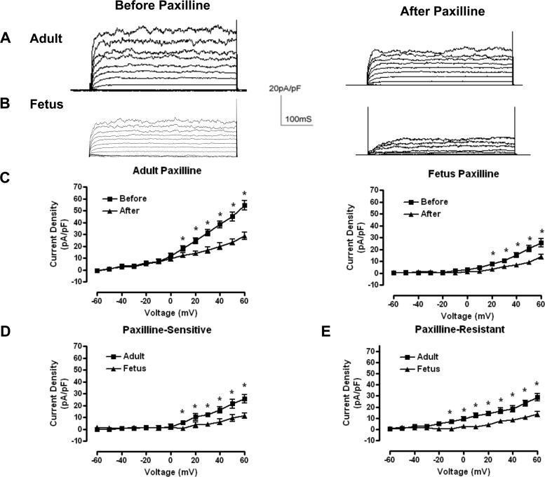
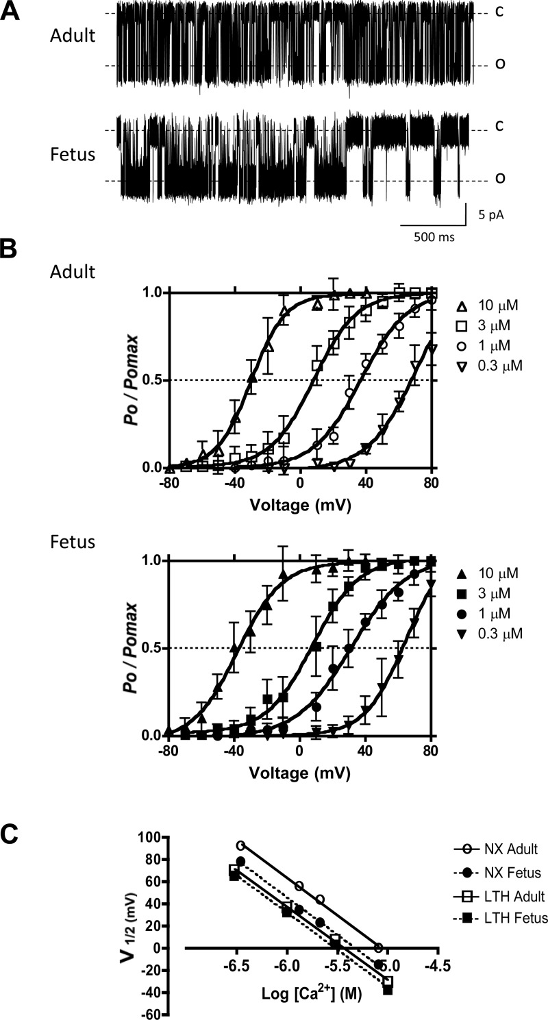
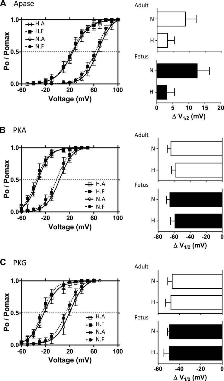
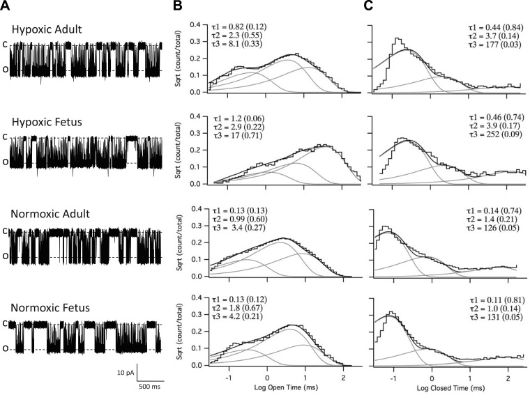
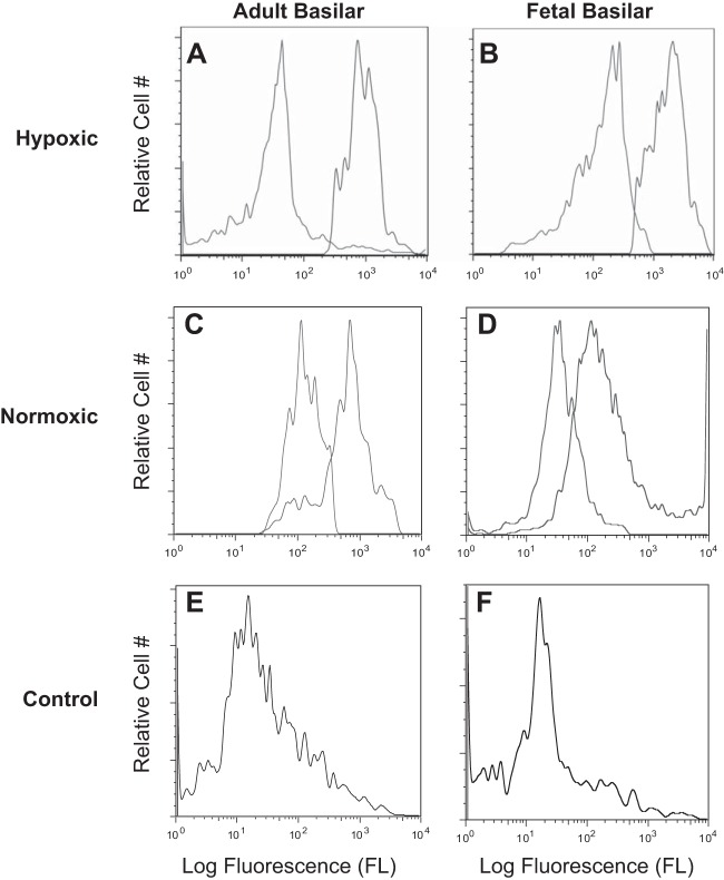
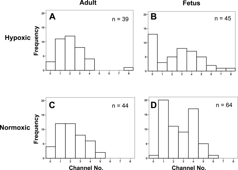
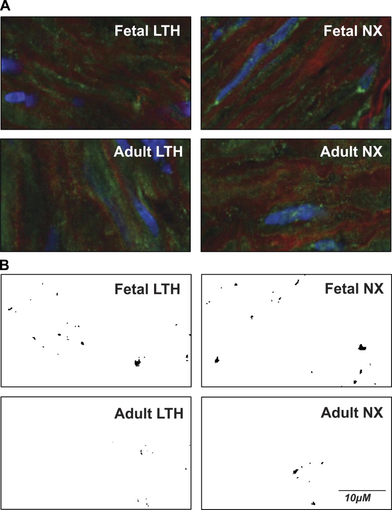
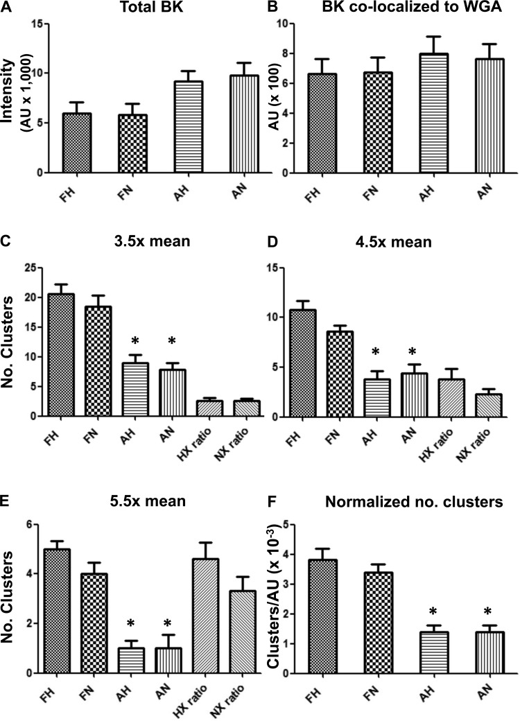
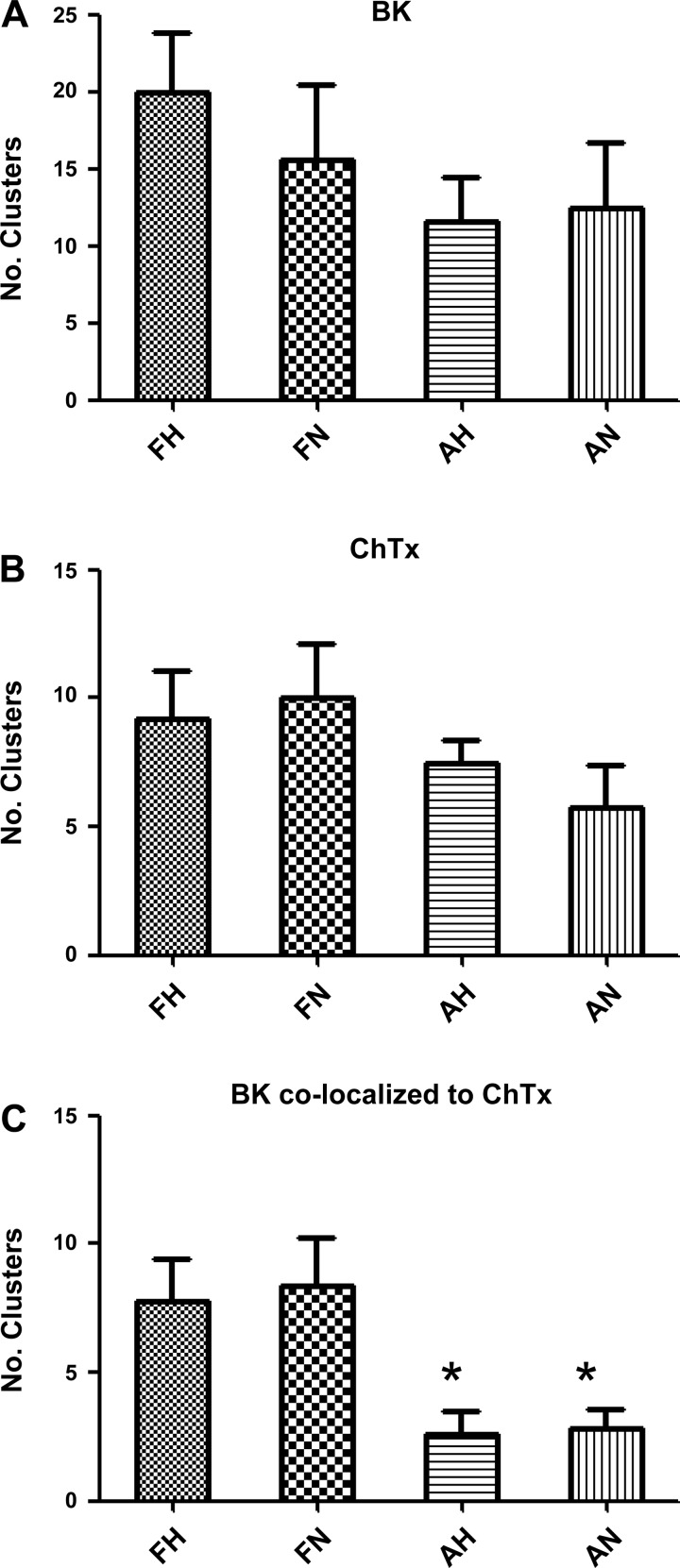
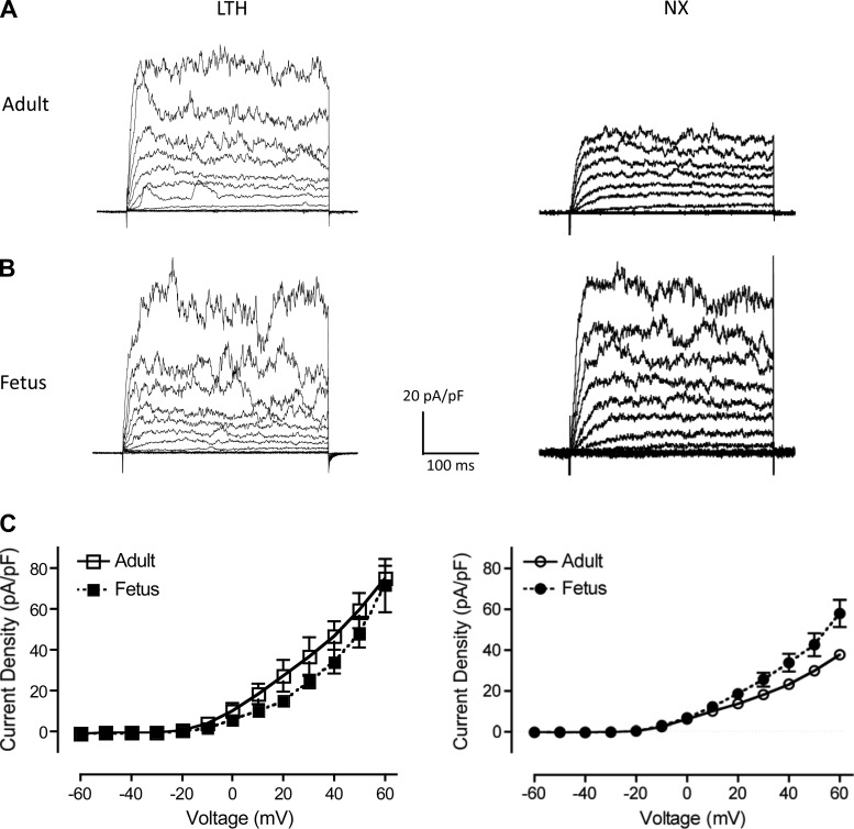
Acclimatization to high-altitude, long-term hypoxia (LTH) reportedly alters cerebral artery contraction-relaxation responses associated with changes in K(+) channel activity. We hypothesized that to maintain oxygenation during LTH, basilar arteries (BA) in the ovine adult and near-term fetus would show increased large-conductance Ca(2+) activated potassium (BK) channel activity. We measured BK channel activity, expression, and cell surface distribution by use of patch-clamp electrophysiology, flow cytometry, and confocal microscopy, respectively, in myocytes from normoxic control and LTH adult and near-term fetus BA. Electrophysiological data showed that BK channels in LTH myocytes exhibited 1) lowered Ca(2+) set points, 2) left-shifted activation voltages, and 3) longer dwell times. BK channels in LTH myocytes also appeared to be more dephosphorylated. These differences collectively make LTH BK channels more sensitive to activation. Studies using flow cytometry showed that the LTH fetus exhibited increased BK β1 subunit surface expression. In addition, in both fetal groups confocal microscopy revealed increased BK channel clustering and colocalization to myocyte lipid rafts. We conclude that increased BK channel activity in LTH BA occurred in association with increased channel affinity for Ca(2+) and left-shifted voltage activation. Increased cerebrovascular BK channel activity may be a mechanism by which LTH adult and near-term fetal sheep can acclimatize to long-term high altitude hypoxia. Our findings suggest that increasing BK channel activity in cerebral myocytes may be a therapeutic target to ameliorate the adverse effects of high altitude in adults or of intrauterine hypoxia in the fetus.

Keywords: Ca2+ signaling; development; high altitude.


Figures

Fig. 1.
Fig. 1.
Whole-cell currents from long-term hypoxia (LTH) adult and fetal smooth muscle cells. A and B: representative whole-cell outward membrane current density traces elicited by a series of 10-mV depolarizing steps (−60 to +60 mV) from a holding potential of −60 mV. Traces before (left) and after (right) paxilline application are shown in typical isolated LTH adult (A) and fetal (B) basilar artery myocytes. Whole-cell current density is obtained from normalized whole-cell currents to membrane capacitance to account for size differences between adult and fetal myocytes. C: averaged steady-state current-voltage plot of outward current density in myocytes obtained from LTH adult (n = 6) and fetal (n = 7) basilar arteries before and after treatment with 5 × 10⁻⁷ M paxilline. D and E: averaged steady-state paxilline-sensitive large-conductance Ca2+-activated K+ channel (BK) currents (left) and residual, paxilline-insensitive currents (right) obtained from digital subtraction of the individual traces such as in A and B. *Significant difference with P < 0.05.
Fig. 2.
Fig. 2.
BK channel open probabilities and calcium set points. A: representative inside-out patch recordings of BK channel from LTH adult and fetal smooth muscle cells in symmetrical 140 mM KCl. In both cases, command potentials were −30 mV making BK channels experience +30 mV depolarizations. The bath [Ca2+] was 1.0 μM. Dotted lines represent the channels in closed (C) and open (O) states. B: voltage activation curves at different membrane potentials in 10-mV increments for various [Ca2+]i. Data are channel open probability (Po) expressed relative to maximum channel open probability (Pomax). Solid lines indicate best-fit curves to the Boltzmann equation: Po/Pomax = 1{1 + exp[(V1/2Vm)/K]}, where V1/2 is the membrane potential (Vm) required for half-maximal activation of the channel and K is the logarithmic voltage sensitivity (change in voltage required for an e-fold increase in activity). The voltage sensitivities estimated from the fitted curves were similar for all concentrations of Ca2+ tested and indicated that channel activity increased e-fold (∼2.72 times) for 23.5 ± 1.8 mV (n = 4, adult) and 25.0 ± 2.1 mV (n = 4, fetus) depolarizations. C: estimation of changes in V1/2 for a 10-fold change in [Ca2+]iV1/2) and estimation of the Ca2+ axis intercept (calcium set point, Ca0) for both adult and fetal BK channels. V1/2 values were obtained from B. The lines represent the best linear regression fits. LTH adult and fetal Ca0 values were calculated to be 3.6 μM and 3.0 μM, respectively. NX, normoxic.
Fig. 3.
Fig. 3.
Effects of exogenous phosphorylation and dephosphorylation on BK channel activities. Single BK channel recordings of BK channels from inside-out micro patches were obtained in 3 μM free [Ca2+] from the isolated myocytes of the 4 experimental animal groups: LTH (H) and normoxic (N) and adult (A) and fetus (F). A: voltage-activation curves of BK channels with alkaline phosphatase (Apase, 350 U/ml) in the bathing medium. Bar graphs summarize the extent to which Apase treatment right-shifts the activation curves in terms of change in V1/2 values for adult (top) and fetal (bottom) groups. B: voltage activation curves of BK channels in the presence of exogenous PKA. After phosphatase pretreatment, purified PKA catalytic subunit (cPKA, 30 U/ml) was added in the presence of KT5823, OA, and ATP. The extent to which PKA left-shifts V1/2 values is summarized on the bar graphs. C: voltage activation curves of BK channels in the presence of exogenous PKG. Following phosphatase pretreatment, purified PKG (2,000 U/ml) was added in the presence of KT5720, OA, and ATP. Effect of PKG left-shift V1/2 values is summarized on the right bar graphs. Solid lines show the best-fit curves to the Boltzmann equation from which V1/2 values were calculated.
Fig. 4.
Fig. 4.
BK channel dwell time analysis. A: representative inside-out patch recordings of BK channels from hypoxic (LTH) adult and fetal, and normoxic adult and fetal myocytes in symmetrical 140 mM KCl solutions with 3 μM free Ca2+. Recordings were done at +60 mV depolarizing potential. C, closed state; O, open state. B and C: plots of open and closed dwell times. Channel open and closed dwell times were plotted on a logarithmic time abscissa as a function of the square root (Sqrt) of the number of events per bin on the ordinate axis. The bin density is 50 bins per decade. Both the open (B) and closed (C) plots were best fitted to exponential functions with 3 components using QuB software (see methods). The lines for the sum and each component exponential fit are shown. The time constants (τ) and their relative weight contributions (in parentheses) of each component to the composite fit are listed.
Fig. 5.
Fig. 5.
Representative flow cytometric distributions of cell surface BK channel βl subunit. A–D: isolated, intact basilar artery smooth myocytes were treated with either primary anti-BK βl (black trace) plus secondary antibody or with secondary antibody alone (gray trace). E and F: primary anti-BK βl antibody was pre-incubated with 70-fold molar excess βl epitopic peptide overnight on ice. Isolated, intact basilar artery smooth myocytes then were treated with the primary antibody and peptide mixture followed by secondary antibody to serve as antibody specificity controls (gray trace). A: LTH adult (n = 8); B: LTH near-term fetus (n = 9); C: normoxic adult (n = 13); D: normoxic near-term fetus (n = 13); E: normoxic adult (n = 13); and F: normoxic fetus (n = 13).
Fig. 6.
Fig. 6.
Number (No.) of BK channels in excised micro patches. The number of BK channels in inside/out patches was determined at +60 mV in symmetrical KCl solutions with 3 mM Ca2+ in the bath solution to ensure maximal channel activation. Patch electrode tip resistances averaged 15.5 ± 0.1 MΩ (n = 192). Frequency histograms of the number of BK channels per patch preparation with distribution curve overlays were displayed. A: LTH adult (n = 39); B: LTH near-term fetus (n = 45); C: normoxic adult (n = 44); and D: normoxic near-term fetus (n = 64).
Fig. 7.
Fig. 7.
Representative confocal microscopic images of arterial myocytes reveal presence of dispersed and clustered BK channels. A: representative color images from adult LTH, fetal LTH, adult NX (normoxic), and fetal NX. Viewed areas measure 20 × 40 μm. Green color indicates presence of BK channels. B: green channel (BK fluorescence) intensities converted to binary image from same areas as above (A) after masking out all values below threshold (3.5× mean intensity). BK clusters show as black areas of different size and shape. Controls with secondary antibody alone or with primary antibody pre-absorbed with antigenic peptide revealed little to no detectable BKα fluorescence (data not shown).
Fig. 8.
Fig. 8.
Total BK channel density, BK surface density, and BK clustering measured in confocal images of intact basilar artery myocytes. A: total BK fluorescence intensity in arbitrary units (AU; means ± SE; n = 5), where FH is fetal hypoxic (LTH), FN is fetal normoxic, AH is adult hypoxic, and AN is adult normoxic. B: BK colocalized with the surrogate surface membrane marker, wheat germ agglutinin (WGA; n = 6). C: number (No.) of BK clusters measured at 3.5 times above mean intensity (n = 7). D: number of BK clusters measured at 4.5 times above mean intensity (n = 6). E: number of BK clusters measured at 5.5 times above mean intensity (n = 6). F: number of BK clusters at 3.5 times mean intensity per total BK intensity (n = 6). Imaged areas measured 20 × 40 μm. Number of animals in each group was either 3 or 4. *Significant difference with P < 0.001 relative to either fetal group. HX ratio and NX ratio in C, D, and E refer to FH:AH and FN:AN, respectively.
Fig. 9.
Fig. 9.
BK channel clusters colocalized to cholera toxin clusters. A: number of BK clusters measured at 3.5 times above mean intensity (means ± SE; n = 5), where FH is fetal hypoxic (LTH), FN is fetal normoxic, AH is adult hypoxic, and AN is adult normoxic. B: number of cholera toxin (ChTx) clusters measured at 3.0 times above mean intensity (n = 5). C: number BK clusters colocalized with ChTx clusters. Imaged areas measured 20 × 40 μm. Number of animals in each group was 3. *Significant difference with P < 0.05 relative to either fetal group.
Fig. 10.
Fig. 10.
Perforated-patch, whole-cell outward current density recordings. A and B: representative whole-cell outward membrane current density traces are shown from isolated LTH and normoxic (NX) adult (A) and fetal (B) basilar artery myocytes. Currents were elicited by a series of 10-mV depolarizing steps (−60 to +60 mV) from a holding potential of −60 mV. Whole-cell current density was used to normalize whole cell currents for size differences between adult and fetal myocytes. C: averaged steady-state current-voltage plot of outward current density in myocytes obtained from LTH (left) adult (n = 5) and fetal (n = 6) and normoxic (right; taken from Ref. 3) adult (n = 4) and fetal (n = 5) basilar arteries.

Similar articles

Cited by

References

    1. Ainslie PN, Subudhi AW. Cerebral blood flow at high altitude. High Alt Med Biol 15: 133–140, 2014. - PubMed
    1. Alioua A, Lu R, Kumar Y, Eghbali M, Kundu P, Toro L, Stefani E. Slo 1 caveolin-binding motif, a mechanism of caveolin-Slo 1 interaction regulating Slo 1 surface expression. J Biol Chem 283: 4809–4817, 2008. - PubMed
    1. Amberg GC, Bonev AD, Rossow CF, Nelson MT, Santana LF. Modulation of the molecular composition of large conductance, Ca2+ activated K+ channels in vascular smooth muscle during hypertension. J Clin Invest 112: 717–724, 2003. - PMC - PubMed
    1. Behrens R, Nolting A, Reimann F, Schwarz M, Waldschutz R, Pongs O. hKCNMB3 and hKCNMB4, cloning and characterization of two members of the large-conductance calcium-activated potassium channel beta subunit family. FEBS Lett 474: 99–106, 2000. - PubMed
    1. Berkefeld H, Fakler B, Schulte U. Ca2+-activated K+ channels: from protein complexes to function. Physiol Rev 90: 1437–1459, 2009. - PubMed

Publication types

MeSH terms

Substances

LinkOut - more resources