. 2015 Apr:76:126-136.
doi: 10.1016/j.nbd.2014.12.032. Epub 2015 Jan 17.

Therapeutic window for cyclooxygenase-2 related anti-inflammatory therapy after status epilepticus

Affiliations

Therapeutic window for cyclooxygenase-2 related anti-inflammatory therapy after status epilepticus

Jianxiong Jiang et al. Neurobiol Dis. 2015 Apr.

Abstract

As a prominent inflammatory effector of cyclooxygenase-2 (COX-2), prostaglandin E2 (PGE2) mediates brain inflammation and injury in many chronic central nervous system (CNS) conditions including seizures and epilepsy, largely through its receptor subtype EP2. However, EP2 receptor activation might also be neuroprotective in models of excitotoxicity and ischemia. These seemingly incongruent observations expose the delicacy of immune and inflammatory signaling in the brain; thus the therapeutic window for quelling neuroinflammation might vary with injury type and target molecule. Here, we identify a therapeutic window for EP2 antagonism to reduce delayed mortality and functional morbidity after status epilepticus (SE) in mice. Importantly, treatment must be delayed relative to SE onset to be effective, a finding that could be explained by the time-course of COX-2 induction after SE and compound pharmacokinetics. A large number of inflammatory mediators were upregulated in hippocampus after SE with COX-2 and IL-1β temporally leading many others. Thus, EP2 antagonism represents a novel anti-inflammatory strategy to treat SE with a tightly-regulated therapeutic window.

Keywords: Chemokine; Cyclooxygenase; Cytokine; EP2; Epilepsy; Gliosis; Interleukin-1β; Neuroinflammation; Prostaglandin; Seizure; Status epilepticus.


Figures

Figure 1
Figure 1. Schematic of seizure induction and drug treatment paradigm
(A) Chemical structure of TG6-10-1. (B) Mice were pretreated with methylscopolamine (2 mg/kg, i.p.) and terbutaline (2 mg/kg, i.p.), and 30 min later injected with pilocarpine (280 mg/kg, i.p.) to induce SE. The SE was allowed to persist for 60 min and terminated by pentobarbital (30 mg/kg, i.p.). Then mice were then randomly split into 4 groups and treated with vehicle or EP2 antagonist TG6-10-1 (5 mg/kg, i.p.) at different time points as indicated. The mice were checked daily for body weight, mortality and nesting behavior. (C) After pilocarpine injection, the behavioral seizure score was tabulated every 5 min until the seizure was terminated by pentobarbital injection 1 h after SE onset (n = 9–28 mice per group). Data are shown as mean ± SEM. (D) The latency to reach behavioral SE after pilocarpine injection (n = 9–28 mice per group). Data are shown as mean + SEM.
Figure 2
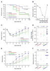
Figure 2. Therapeutic window of blocking EP2 receptor to treat status epilepticus (SE)
(A) Survival rates of animals that received vehicle or TG6-10-1 treatment (n = 9–28 mice per group) for up to 60 d after SE (P = 0.008 for delayed treatment 2 compared with vehicle group, Kaplan-Meier survival analysis; P = 0.029 for delayed treatment 1 as reported by Jiang et al. 2013). (B) Effect of the first dosing time after SE onset on animal 60-day delayed mortality. (C) Effect of TG6-10-1 on mouse body weight change after SE (n = 9–28 mice per time point, *P < 0.05; **P < 0.01; ***P < 0.001 compared with vehicle group, two-way ANOVA with post-hoc Bonferroni test). Data are shown as mean ± SEM. (D) Body weight change of individual animals from day 1 to day 4 after SE (n = 7–22 mice per group, *P = 0.045, Fisher’s exact test). The average weight changes from day 1 to day 4 after SE for each group are indicated by bars. (E) Effect of TG6-10-1 on mouse nesting behavior after SE (n = 9–28 mice per time point, **P < 0.01; ***P < 0.001 compared with vehicle group, two-way ANOVA with post-hoc Bonferroni test). Data are shown as mean ± SEM. (F) Nesting score of individual animals at day 4 after SE (n = 7–22 mice per group, ***P = 0.0003, Fisher’s exact test). The average nesting scores at day 4 after SE for each group are indicated by bars.
Figure 3
Figure 3. Inflammatory genes are upregulated following pilocarpine-induced SE
Quantitative real-time PCR (qRT-PCR) was performed to measure the time-course of changes in mRNA levels of a number of inflammatory mediators and markers in mouse hippocampi after pilocarpine-induced SE. These molecules include enzymes: COX-2, mPGES-1, iNOS and NOX-2 (gp91phox); cytokines: IL-1β, IL-6, TNF-α and TGF-β1; chemokines: CCL2, CCL3 and CCL4; EP2 receptor; and gliosis markers: GFAP, S100B, Iba1 and CD11b. All tested genes were substantially induced by SE with COX-2 and IL-1β temporally leading many others (n = 6–8 mice per time point, *P < 0.05; **P < 0.01 compared with control mice, one-way ANOVA and post-hoc Dunnett’s test of the ΔΔCT values from the qRT-PCR). Data are shown as mean ± SEM.
Figure 4
Figure 4. COX-2 and IL-1β proteins are rapidly induced by SE
(A) The COX-2, EP2, IL-1β and GFAP protein levels in mouse hippocampi after SE were measured by western blot analysis with GAPDH as the loading control. Three representative samples from each time point after SE onset are shown on the blots. (B) The blots were scanned and the band intensities were assessed by ImageJ. The relative protein levels of COX-2, EP2, IL-1β (pre-pro, pro-, and mature forms) and GFAP were normalized to their mean basal levels. COX-2, IL-1β and GFAP protein levels, but not EP2, were rapidly and substantially upregulated in mouse hippocampi after pilocarpine-induced SE (n = 6 mice per time point, *P < 0.05; **P < 0.01 compared with control mice, one-way ANOVA and post-hoc Dunnett’s test). Data are shown as mean + SEM.
Figure 5
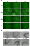
Figure 5. Immunohistochemistry of COX-2 in hippocampus after pilocarpine-induced SE in mice
(A) Immunostaining was performed to examine COX-2 protein expression and location in hippocampal subregions: dentate gyrus (DG), cornu ammonis area 1 (CA1) and CA3, 1 d, 4 d or 10 d after pilocarpine-induced SE in mice, visualized by fluorescence. Scale bar = 100 μm. (B) Time-course expression of COX-2 protein in mouse hippocampal CA3 subregion, 1 h, 4 h, 12 h, 1 d or 3 d after SE onset, assessed by 3,3′-diaminobenzidine (DAB) staining. Note that SE induced COX-2 protein expression in hippocampal principal neurons, beginning within 1 h after SE onset. The COX-2 protein induction peaked by 4 to 12 h, and subsided by 3 d after SE. Scale bar = 100 μm.
Figure 6
Figure 6. EP2 receptor mediates expression of pro-inflammatory genes after SE
The mRNA levels of an array of pro-inflammatory mediators or markers were measured by qRT-PCR in mouse hippocampi after pilocarpine SE, including oxidative stress-related enzymes: COX-2, mPGES-1; iNOS, NOX-2; pro-inflammatory cytokines: IL-1β, IL-6, TNF-α, TGF-β1, CCL2, CCL3, CCL4; and gliosis markers: GFAP, S100B, Iba1, CD11b. mRNA induction of each gene was shown by comparing with its basal level in mice not treated with pilocarpine. (A) Administration of EP2 antagonist TG6-10-1 (at 4 and 21 h after SE onset) had no effect on mRNA induction of most pro-inflammatory genes, measured 1 d after SE (n = 5–6 mice per group, P = 0.216, two-tailed paired t test). Data are shown as mean + SEM. (B) Administration of EP2 antagonist TG6-10-1 (at 4, 21 and 30 h after SE onset) significantly reduced SE-triggered pro-inflammatory gene induction, measured 4 d after SE (n = 8 mice per group, P = 0.036, two-tailed paired t test). Data are shown as mean + SEM.

References

    1. Andreasson K. Emerging roles of PGE2 receptors in models of neurological disease. Prostaglandins Other Lipid Mediat. 2010;91:104–12. - PMC - PubMed
    1. Aronica E, Crino PB. Inflammation in epilepsy: clinical observations. Epilepsia. 2011;52(Suppl 3):26–32. - PubMed
    1. Avignone E, Ulmann L, Levavasseur F, Rassendren F, Audinat E. Status epilepticus induces a particular microglial activation state characterized by enhanced purinergic signaling. J Neurosci. 2008;28:9133–44. - PMC - PubMed
    1. Balosso S, Ravizza T, Aronica E, Vezzani A. The dual role of TNF-alpha and its receptors in seizures. Exp Neurol. 2013;247:267–71. - PubMed
    1. Bazan NG. COX-2 as a multifunctional neuronal modulator. Nat Med. 2001;7:414–5. - PubMed

Publication types

MeSH terms