Review
. 2015 Feb;40(2):101-7.
doi: 10.1016/j.tibs.2014.12.006. Epub 2015 Jan 16.

Insights into the trimeric HIV-1 envelope glycoprotein structure

Affiliations
Review

Insights into the trimeric HIV-1 envelope glycoprotein structure

Andrew B Ward et al. Trends Biochem Sci. 2015 Feb.

Abstract

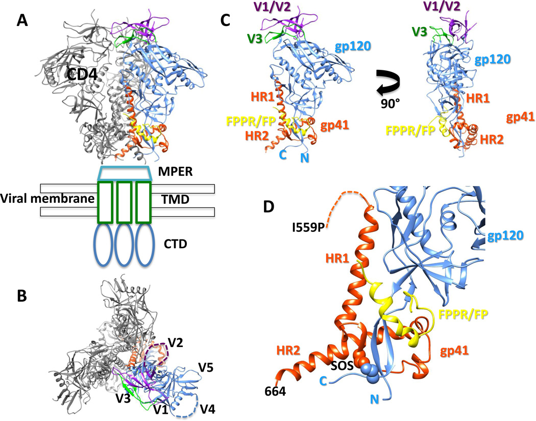
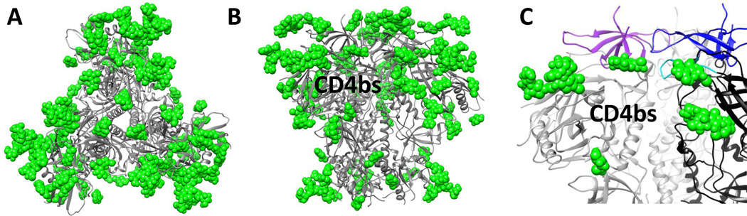
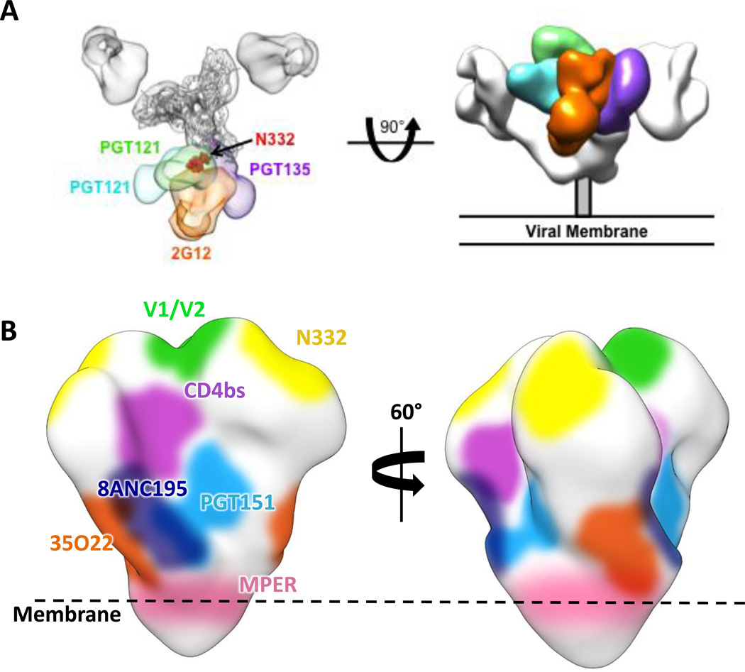
The HIV-1 envelope glycoprotein (Env) trimer is responsible for receptor recognition and viral fusion with CD4(+) T cells, and is the sole target for neutralizing antibodies. Thus, understanding its molecular architecture is of significant interest. However, the Env trimer has proved to be a challenging target for 3D structure determination. Recent electron microscopy (EM) and X-ray structures have at last enabled us to decipher the structural complexity and unique features of the Env trimer, and how it is recognized by an ever-expanding arsenal of potent broadly neutralizing antibodies. We describe our current knowledge of the Env trimer structure in the context of exciting recent developments in the identification and characterization of HIV broadly neutralizing antibodies.

Keywords: HIV-1; broadly neutralizing antibodies; envelope glycoprotein trimer; structure.


Figures

Fig. 1
Fig. 1
Structure of the soluble BG505 SOSIP.664 Env trimer. (A) Side and (B) top views of Env trimer (PDB 4TVP). The receptor CD4 binding site is labeled CD4. Structures of the membrane proximal external region (MPER), transmembrane domain (TMD) and cytoplasmic domain (CTD) have not yet been determined. The variable loops (V1-V5) are located at the apex of the trimer and project outward. V2 and V4 (dashed lines) are disordered in the current structures. (C) A single gp120/gp41 protomer is displayed. The gp41 components heptad repeat 1 and 2 (HR1, HR2) are positioned at the base of the trimer. The fusion peptide proximal region (FPPR) and fusion peptide (FP) are located at the gp120 interface. The N and C termini of gp120 form important interactions with gp41. (D) Close up of gp41. The stabilizing disulfide bond (SOS) introduced at residues 501 (gp120) and 605 (gp41) is shown in spheres. The isoleucine to proline (I559P) is disordered in the current structures and its approximate location is shown with a dashed line. The C terminus of gp41 in the BG505 SOSIP.664 trimer is labeled as 664.
Fig. 2
Fig. 2
The highly glycosylated surface of Env. (A) Top and (B) side views of the Env trimer (gray cartoon) illustrating the extensive glycan shield. Shown in green are the glycans resolved in the current structures of the Env trimer. The full extent of most glycans remains to be determined so the figure here under-represents the extent of glycans. The receptor CD4 binding site is labeled. (C) Close up illustrating the variable loop and glycan constraints restricting access to the CD4 binding site. The V1/V2 regions of two different protomers are colored purple and blue. V3 is colored in cyan. Glycans are displayed as green spheres.
Fig. 3
Fig. 3
Sites of vulnerability on the surface of HIV-1 Env. (A) Top and side view HIV-1 Env trimer bound to different antibodies that all recognize the N332 glycan, but approach from different angles. The antibodies are colored separately and superimposed on the electron microscopy (EM) map EMD-5982. The crystal structure of gp120 (gray cartoon) is docked in the EM map and the N332 glycan is indicated by red spheres. (B) Known epitopes for bnAbs colored on the surface of the EM map EMD-5919.

References

    1. Bartesaghi A, et al. Prefusion structure of trimeric HIV-1 envelope glycoprotein determined by cryo-electron microscopy. Nat. Struct. Mol. Biol. 2013;20:1352–1357. - PMC - PubMed
    1. Binley JM, et al. A recombinant human immunodeficiency virus type 1 envelope glycoprotein complex stabilized by an intermolecular disulfide bond between the gp120 and gp41 subunits is an antigenic mimic of the trimeric virion-associated structure. J. Virol. 2000;74:627–643. - PMC - PubMed
    1. Blattner C, et al. Structural delineation of a quaternary, cleavage-dependent epitope at the gp41-gp120 interface on intact HIV-1 Env trimers. Immunity. 2014;40:669–680. - PMC - PubMed
    1. Bonomelli C, et al. The glycan shield of HIV is predominantly oligomannose independently of production system or viral clade. PLoS One. 2011;6:e23521. - PMC - PubMed
    1. Davenport TM, et al. Isolate-specific differences in the conformational dynamics and antigenicity of HIV-1 gp120. J. Virol. 2013;87:10855–10873. - PMC - PubMed

MeSH terms