Case Reports
. 2015 Aug;43(4):495-501.
doi: 10.1007/s15010-015-0720-y. Epub 2015 Jan 20.

Severe neurologic syndrome associated with Middle East respiratory syndrome corona virus (MERS-CoV)

Affiliations
Case Reports

Severe neurologic syndrome associated with Middle East respiratory syndrome corona virus (MERS-CoV)

Y M Arabi et al. Infection. 2015 Aug.

Abstract

Background: Since the identification of the first case of infection with the Middle East respiratory syndrome corona virus (MERS-CoV) in Saudi Arabia in June 2012, the number of laboratory-confirmed cases has exceeded 941 cases globally, of which 347 died. The disease presents as severe respiratory infection often with shock, acute kidney injury, and coagulopathy. Recently, we observed three cases who presented with neurologic symptoms. These are so far the first reported cases of neurologic injury associated with MERS-CoV infection.

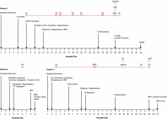
Methods: Data was retrospectively collected from three patients admitted with MERS-CoV infection to Intensive Care unit (ICU) at King Abdulaziz Medical City, Riyadh. They were managed separately in three different wards prior to their admission to ICU.

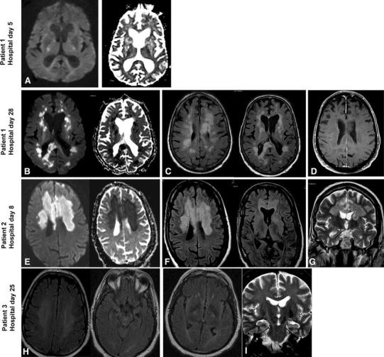
Finding: The three patients presented with severe neurologic syndrome which included altered level of consciousness ranging from confusion to coma, ataxia, and focal motor deficit. Brain MRI revealed striking changes characterized by widespread, bilateral hyperintense lesions on T2-weighted imaging within the white matter and subcortical areas of the frontal, temporal, and parietal lobes, the basal ganglia, and corpus callosum. None of the lesions showed gadolinium enhancement.

Interpretation: CNS involvement should be considered in patients with MERS-CoV and progressive neurological disease, and further elucidation of the pathophysiology of this virus is needed.


Figures

Fig. 1
Fig. 1
Timeline of pertinent events and MERS-CoV RT-PCR results in patients 1, 2, and 3. MRI Magnetic resonance imaging, CCRT critical care response team, ICU intensive care unit, RRT renal replacement therapy, TA tracheal aspirate, BAL bronchoalveolar lavage, CSF cerebrospinal fluid, NSTEMI non-ST elevation myocardial infarction, GI gastrointestinal
Fig. 2
Fig. 2
Magnetic resonance imaging (MRI) of the three patients. Patient 1—First MRI—day 5: a Diffusion weighted imaging (DWI) and Apparent Diffusion Coefficient (ADC) mapping showed patchy non-specific signal changes with no restriction. Further images were not obtained because patient could not tolerate the test. Patient 1—Second MRI—day 28: b Diffusion weighted imaging (DWI) and Apparent Diffusion Coefficient (ADC) mapping showed diffusion restriction of the multiple white matter lesions. c Axial fluid-attenuated inversion recovery (FLAIR) images showed multiple hyperintense lesions in the subcortical areas and deep white matter of frontal, temporal, and parietal lobes bilaterally as well as in the corpus callosum. d Axial contrast enhanced T1-weighted (T1Wl) showed multiple subcortical areas and deep white matter non-enhancing lesions. Patient 2—Day 8: e DWI and ADC mapping showed diffusion restriction of the bilateral frontal and corpus callosum lesions. f Axial FLAIR images showed bilateral confluent subcortical areas and deep white matter hyperintense lesions in the frontal lobes, basal ganglia, and corpus callosum. g Coronal T2 images showed bilateral hyperintense white matter lesions in the frontal lobes and in the corpus callosum. Patient 3—Day 25: h Axial FLAIR Images showing diffuse white matter (deep) signal abnormality at the insular region bilaterally. i Coronal T2-weighted images showing abnormality involving corticospinal tract bilaterally extending to the brain stem

References

    1. Zaki AM, van Boheemen S, Bestebroer TM, Osterhaus AD, Fouchier RA. Isolation of a novel coronavirus from a man with pneumonia in Saudi Arabia. New Engl J Med. 2012;367:1814–1820. doi: 10.1056/NEJMoa1211721. - DOI - PubMed
    1. Middle East respiratory syndrome coronavirus (MERS-CoV)—update. http://www.who.int/csr/don/26-december-2014-mers/en/. Accessed 15 Jan 2015 - PMC - PubMed
    1. The Who Mers-Cov Research G. State of knowledge and data gaps of Middle East Respiratory Syndrome Coronavirus (MERS-CoV) in humans. PLoS Curr. 2013; 5. - PMC - PubMed
    1. Arabi YM, Arifi AA, Balkhy HH, Najm H, Aldawood AS, Ghabashi A, et al. Clinical course and outcomes of critically ill patients with Middle East respiratory syndrome coronavirus infection. Ann Intern Med. 2014;60:389–397. - PubMed
    1. Momattin H, Mohammed K, Zumla A, Memish ZA, Al-Tawfiq JA. Therapeutic options for Middle East respiratory syndrome coronavirus (MERS-CoV)—possible lessons from a systematic review of SARS-CoV therapy. Int J Infect Dis. 2013;17:e792–e798. doi: 10.1016/j.ijid.2013.07.002. - DOI - PMC - PubMed

Publication types

MeSH terms

LinkOut - more resources