. 2015 Jan 20:6:5939.
doi: 10.1038/ncomms6939.

A dynamically coupled allosteric network underlies binding cooperativity in Src kinase

Affiliations

A dynamically coupled allosteric network underlies binding cooperativity in Src kinase

Zachariah H Foda et al. Nat Commun. .

Abstract

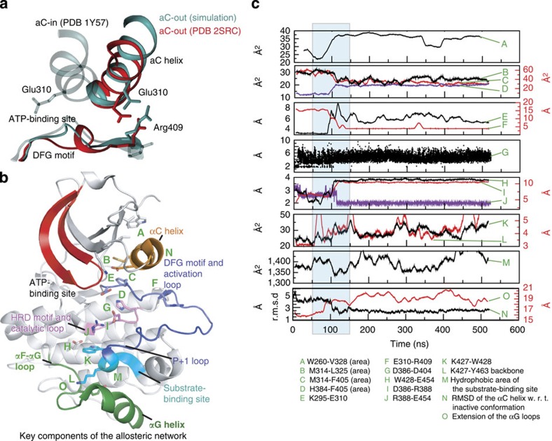
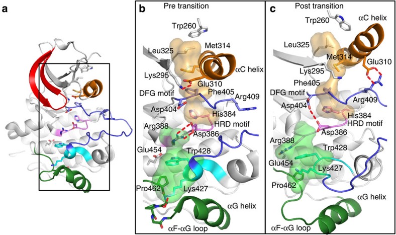
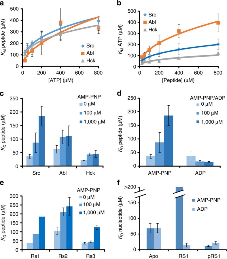
Protein tyrosine kinases are attractive drug targets because many human diseases are associated with the deregulation of kinase activity. However, how the catalytic kinase domain integrates different signals and switches from an active to an inactive conformation remains incompletely understood. Here we identify an allosteric network of dynamically coupled amino acids in Src kinase that connects regulatory sites to the ATP- and substrate-binding sites. Surprisingly, reactants (ATP and peptide substrates) bind with negative cooperativity to Src kinase while products (ADP and phosphopeptide) bind with positive cooperativity. We confirm the molecular details of the signal relay through the allosteric network by biochemical studies. Experiments on two additional protein tyrosine kinases indicate that the allosteric network may be largely conserved among these enzymes. Our work provides new insights into the regulation of protein tyrosine kinases and establishes a potential conduit by which resistance mutations to ATP-competitive kinase inhibitors can affect their activity.


Figures

Figure 1
Figure 1. Concerted conformational change simulated in Src kinase up on protonation.
(a) The movement of the αC helix in the MD simulations. Starting from the active conformation (Simulation—Start), the αC helix (cyan transparent) rotated outwards by about 120° (cyan solid) leading to a salt bridge between Glu310 and Arg409 (Simulation—End). The structure at the end of the simulation resembles that in the crystal structure (red) of autoinhibited Src kinase (Experiment—Inactive, PDB entry 2SRC). (b) The location of the contiguous network of residues involved in the concerted conformational change. Green letters denote the approximate location of conformational changes quantified in c; a more detailed view of the key residues involved in the conformational change is shown in Fig. 2b,c. (c) The structural parameters (contact area, salt bridge or hydrogen bond distance and r.m.s.d.) characterizing the conformational change shown as functions of simulation time. Light blue is used to highlight the narrow time window in which the concerted conformational change occurred.
Figure 2
Figure 2. Atomic details of the concerted conformational change.
(a) An overview of the active Src kinase domain (from PDB entry 1Y57). Helix αC is coloured orange, P-loop red, activation loop blue, catalytic loop pink, P+1 loop cyan and helix αG with αG-αF loop green. Black rectangle indicates the enlarged area to the right. (b,c) Close-up of the residues identified as part of the allosteric network before (b) and after (c) the transition is observed in the simulations. The P-loop and parts of the activation loop have been omitted for clarity. The hydrophobic spine residues (orange transparent surface) and the substrate-binding site (green transparent surface) are highlighted. (b) Prepared using PDB 1Y57, from which the simulation was initiated. (c) Prepared using a snapshot from a simulation, but the key features highlighted (for example, the positions of Trp260 and the Glu310-Arg409 salt bridge) are also seen in the crystal structure of inactive Src kinase (PDB 2SRC). The key hydrogen bond between protonated Asp404 and Asp386 is seen in other kinase crystal structures (for example, PDB 1JOW and 1M14).
Figure 3
Figure 3. Negative cooperativity of ATP and substrate binding and positive cooperativity of ADP and substrate binding.
(a) The effect of ATP concentration on substrate Km for Src, Abl and Hck kinase domains. (b) The effect of substrate peptide concentration on ATP Km for Src, Abl and Hck kinase domains. (c) The effect of AMP-PNP concentration on substrate Kd for Src, Abl and Hck kinase domains. (d) The effect of ADP and AMP-PNP concentration on substrate Kd for the Src kinase domain. (e) Dissociation constants for peptides of different sequences at increasing AMP-PNP concentration. Rs1: Src-optimal substrate peptide (AEEEIYGEFAKKK); Rs2: peptide sequence (AEEMIYGEFAKKK); Rs3: peptide sequence (GIYWHHY). Km values were determined in a kinase activity assay. Kd values for the substrate peptides were determined using fluorescence anisotropy at 1 μM labelled peptide. Kd values for AMP-PNP were determined using isothermal titration calorimetry. (f) The effect of peptide and phosphorylated peptide on ADP and AMP-PNP binding. All experiments were performed in triplicate and data represent mean values±s.e.m.
Figure 4
Figure 4. Mutations to the allosteric network producing biochemical phenotypes.
(a) D404N and D386N mutations leading to stronger substrate binding and disrupting the binding cooperativity, respectively. (b) Effect of W260A on ATP-binding affinity. (c) Effect of W260A on peptide-binding affinity. (d) Effect of T338I on ATP-binding affinity. (e) Effect of T338I on substrate-binding affinity. Kd values were determined using fluorescence anisotropy at 1 μM labelled Src-optimized peptide (Rs1). All experiments were performed in triplicate and data represent mean values±s.e.m.
Figure 5
Figure 5. The allosteric network and negative cooperativity in the context of a kinase catalytic cycle.
(a) The key components of the allosteric network are shown in two configurations. The protonation of the DFG aspartate repositions the Asp and Phe residues, which disrupts the regulatory hydrophobic spine and leads to the αC-out transition and the repositioning of Trp260 in the N-lobe, and in the C-lobe induces the RAA/AAR Arg to form alternative interactions with Trp428 and Glu454, resulting in a rearrangement of the substrate-binding site. (b) In a catalytic cycle, the apo kinase (I) binds ATP/Mg2+ and substrate, yielding the bisubstrate complex (II), in which the phosphoryl transfer from ATP to substrate occurs. Following the phosphoryl-transfer step, the DFG aspartate becomes protonated (III). The phosphorylated substrate is subsequently released (IV), which weakens ADP binding through the cooperative mechanism and promotes ADP release. The DFG aspartate is then once again deprotonated, and the affinity for ATP increases, starting the catalytic cycle again (I).

References

    1. Schlessinger J. Cell signaling by receptor tyrosine kinases. Cell 103, 211–225 (2000). - PubMed
    1. Rubin G. M. et al.. Comparative genomics of the eukaryotes. Science 287, 2204–2215 (2000). - PMC - PubMed
    1. Manning G., Whyte D. B., Martinez R., Hunter T. & Sudarsanam S. The protein kinase complement of the human genome. Science 298, 1912–1934 (2002). - PubMed
    1. Hagerkvist R., Makeeva N., Elliman S. & Welsh N. Imatinib mesylate (Gleevec) protects against streptozotocin-induced diabetes and islet cell death in vitro. Cell Biol. Int. 30, 1013–1017 (2006). - PubMed
    1. Vlahovic G. & Crawford J. Activation of tyrosine kinases in cancer. Oncologist 8, 531–538 (2003). - PubMed

Publication types

MeSH terms

Substances