

Enhancement of fear extinction with deep brain stimulation: evidence for medial orbitofrontal involvement
- PMID: 25601229
- PMCID: PMC4915256
- DOI: 10.1038/npp.2015.20
Enhancement of fear extinction with deep brain stimulation: evidence for medial orbitofrontal involvement
Abstract
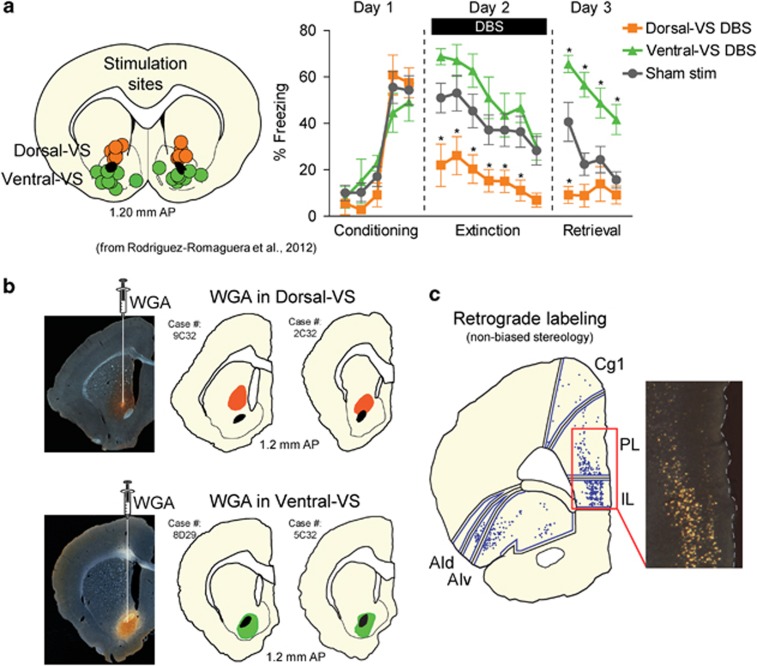
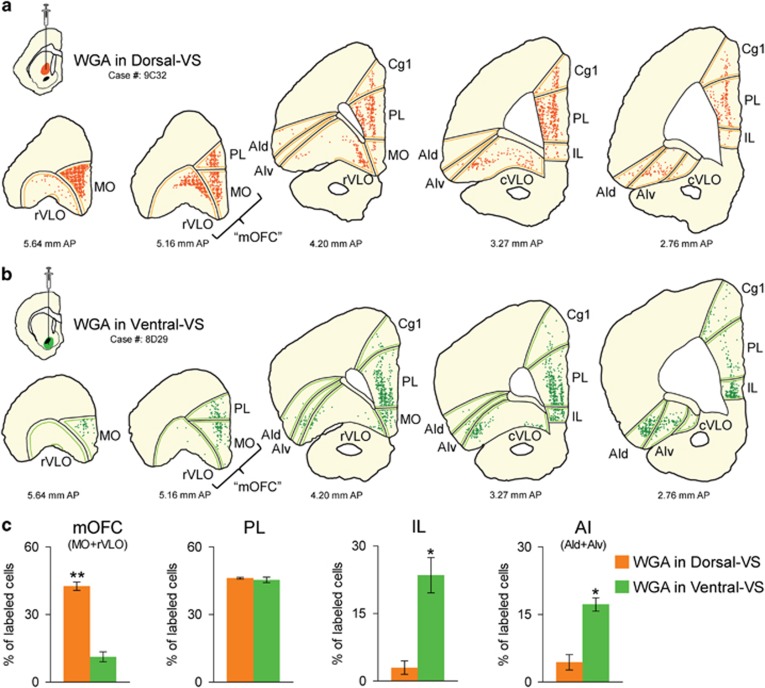
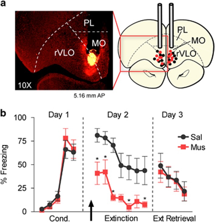
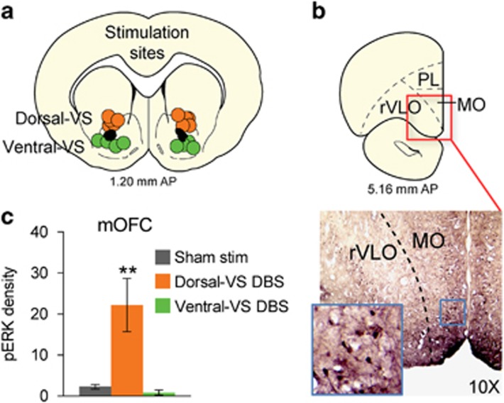
Deep brain stimulation (DBS) of the ventral capsule/ventral striatum (VC/VS) reduces anxiety, fear, and compulsive symptoms in patients suffering from refractory obsessive-compulsive disorder. In a rodent model, DBS-like high-frequency stimulation of VS can either enhance or impair extinction of conditioned fear, depending on the location of electrodes within VS (dorsal vs ventral). As striatal DBS activates fibers descending from the cortex, we reasoned that the differing effects on extinction may reflect differences in cortical sources of fibers passing through dorsal-VS and ventral-VS. In agreement with prior anatomical studies, we found that infralimbic (IL) and anterior insular (AI) cortices project densely through ventral-VS, the site where DBS impaired extinction. Contrary to IL and AI, we found that medial orbitofrontal cortex (mOFC) projects densely through dorsal-VS, the site where DBS enhanced extinction. Furthermore, pharmacological inactivation of mOFC reduced conditioned fear and DBS of dorsal-VS-induced plasticity (pERK) in mOFC neurons. Our results support the idea that VS DBS modulates fear extinction by stimulating specific fibers descending from mOFC and prefrontal cortices.
Figures
Similar articles
-
Deep brain stimulation of the ventral striatum enhances extinction of conditioned fear.Proc Natl Acad Sci U S A. 2012 May 29;109(22):8764-9. doi: 10.1073/pnas.1200782109. Epub 2012 May 14. Proc Natl Acad Sci U S A. 2012. PMID: 22586125 Free PMC article.
-
Bidirectional Modulation of Extinction of Drug Seeking by Deep Brain Stimulation of the Ventral Striatum.Biol Psychiatry. 2016 Nov 1;80(9):682-690. doi: 10.1016/j.biopsych.2016.05.015. Epub 2016 May 27. Biol Psychiatry. 2016. PMID: 27449798 Free PMC article.
-
Dissociable roles of prelimbic and infralimbic cortices, ventral hippocampus, and basolateral amygdala in the expression and extinction of conditioned fear.Neuropsychopharmacology. 2011 Jan;36(2):529-38. doi: 10.1038/npp.2010.184. Epub 2010 Oct 20. Neuropsychopharmacology. 2011. PMID: 20962768 Free PMC article.
-
Deep Brain Stimulation for Refractory Obsessive-Compulsive Disorder: Towards an Individualized Approach.Front Psychiatry. 2019 Dec 13;10:905. doi: 10.3389/fpsyt.2019.00905. eCollection 2019. Front Psychiatry. 2019. PMID: 31920754 Free PMC article. Review.
-
Prefrontal mechanisms in extinction of conditioned fear.Biol Psychiatry. 2006 Aug 15;60(4):337-43. doi: 10.1016/j.biopsych.2006.03.010. Epub 2006 May 19. Biol Psychiatry. 2006. PMID: 16712801 Review.
Cited by
-
Prefrontal cortical BDNF: A regulatory key in cocaine- and food-reinforced behaviors.Neurobiol Dis. 2016 Jul;91:326-35. doi: 10.1016/j.nbd.2016.02.021. Epub 2016 Feb 26. Neurobiol Dis. 2016. PMID: 26923993 Free PMC article. Review.
-
It takes two: Bilateral bed nuclei of the stria terminalis mediate the expression of contextual fear, but not of moderate cued fear.Prog Neuropsychopharmacol Biol Psychiatry. 2020 Jul 13;101:109920. doi: 10.1016/j.pnpbp.2020.109920. Epub 2020 Mar 10. Prog Neuropsychopharmacol Biol Psychiatry. 2020. PMID: 32169558 Free PMC article.
-
Deep Brain Stimulation in Animal Models of Fear, Anxiety, and Posttraumatic Stress Disorder.Neuropsychopharmacology. 2016 Nov;41(12):2810-2817. doi: 10.1038/npp.2016.34. Epub 2016 Mar 2. Neuropsychopharmacology. 2016. PMID: 26932819 Free PMC article. Review.
-
Mapping structural covariance networks in children and adolescents with post-traumatic stress disorder after earthquake.Front Psychiatry. 2022 Sep 15;13:923572. doi: 10.3389/fpsyt.2022.923572. eCollection 2022. Front Psychiatry. 2022. PMID: 36186852 Free PMC article.
-
The prefrontal cortex and OCD.Neuropsychopharmacology. 2022 Jan;47(1):211-224. doi: 10.1038/s41386-021-01130-2. Epub 2021 Aug 16. Neuropsychopharmacology. 2022. PMID: 34400778 Free PMC article. Review.
References
-
- Baxter LR Jr., Saxena S, Brody AL, Ackermann RF, Colgan M, Schwartz JM et al (1996). Brain mediation of obsessive-compulsive disorder symptoms: evidence from functional brain imaging studies in the human and nonhuman primate. Semin Clin Neuropsychiatry 1: 32–47. - PubMed
-
- Berendse HW, Galis-de Graaf Y, Groenewegen HJ (1992). Topographical organization and relationship with ventral striatal compartments of prefrontal corticostriatal projections in the rat. J Comp Neurol 316: 314–347. - PubMed
-
- Burgos-Robles A, Vidal-Gonzalez I, Santini E, Quirk GJ (2007). Consolidation of fear extinction requires NMDA receptor-dependent bursting in the ventromedial prefrontal cortex. Neuron 53: 871–880. - PubMed
Publication types
MeSH terms
Substances
Grants and funding
LinkOut - more resources
Full Text Sources
Other Literature Sources
Research Materials
