. 2015 Apr;96(1):163-74.
doi: 10.1111/mmi.12930. Epub 2015 Feb 11.

Oxidative stress conditions increase the frequency of de novo formation of the yeast [PSI+] prion

Affiliations

Oxidative stress conditions increase the frequency of de novo formation of the yeast [PSI+] prion

Victoria A Doronina et al. Mol Microbiol. 2015 Apr.

Abstract

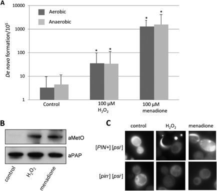
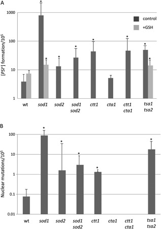
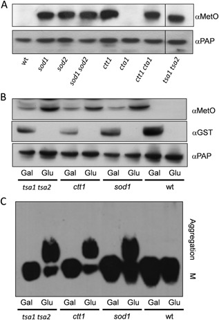
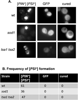
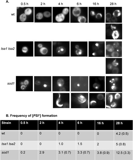
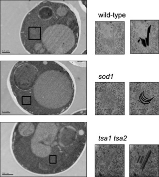
Prions are self-perpetuating amyloid protein aggregates which underlie various neurodegenerative diseases in mammals and heritable traits in yeast. The molecular basis of how yeast and mammalian prions form spontaneously into infectious amyloid-like structures is poorly understood. We have explored the hypothesis that oxidative stress is a general trigger for prion formation using the yeast [PSI(+)] prion, which is the altered conformation of the Sup35 translation termination factor. We show that the frequency of [PSI(+)] prion formation is elevated under conditions of oxidative stress and in mutants lacking key antioxidants. We detect increased oxidation of Sup35 methionine residues in antioxidant mutants and show that overexpression of methionine sulphoxide reductase abrogates both the oxidation of Sup35 and its conversion to the [PSI(+)] prion. [PSI(+)] prion formation is particularly elevated in a mutant lacking the Sod1 Cu,Zn-superoxide dismutase. We have used fluorescence microscopy to show that the de novo appearance of [PSI(+)] is both rapid and increased in frequency in this mutant. Finally, electron microscopy analysis of native Sup35 reveals that similar fibrillar structures are formed in both the wild-type and antioxidant mutants. Together, our data indicate that oxidative stress is a general trigger of [PSI(+) formation, which can be alleviated by antioxidant defenses.


Figures

Figure 1
Figure 1
Oxidative stress conditions increase the frequency of [PSI +] prion formation. A. [PSI +] prion formation was quantified using an engineered ura3–14 allele, which contains the ade1–14 nonsense mutation inserted into the wild‐type URA 3 gene (Manogaran et al., 2006). [PSI +] prion formation was scored by growth on media lacking uracil, indicative of decreased translational termination efficiency. [PSI +] formation was also scored by growth on media lacking uracil under anaerobic conditions. [PSI +] formation was differentiated from nuclear gene mutations which give rise to uracil prototrophy by their irreversible elimination in GdnHCl. The control [PIN +] [psi ] strain was grown in the presence of 100 μM hydrogen peroxide or 100 μM menadione for 16 h prior to scoring [PSI +] prion formation (in the absence of exogenous oxidants). Data shown are the means of at least three independent experiments expressed as the frequency of [PSI +] prion colonies per 105 viable cells. Error bars denote the standard deviation. *P < 0.05. B. Western blot analysis of Sup35 oxidation. Sup35 was affinity purified using TAP chromatography from a control [PIN +] [psi ] strain following growth in the presence of 100 μM hydrogen peroxide or 100 μM menadione for 16 h. Western blots were probed with α‐PAP to confirm that similar amounts of Sup35 were purified from each strain. Sup35 oxidation was detected using antibodies that recognize methionine sulphoxide (αMetO). C. Representative fluorescence micrographs are shown for the [PIN +] [psi +] control strain containing the Sup35NMGFP plasmid following growth in the presence of 100 μM hydrogen peroxide or 100 μM menadione for 16 h. The Sup35NMGFP plasmid was induced for 2 h using copper prior to visualizing aggregate formation.
Figure 2
Figure 2
The frequency of [PSI +] formation is increased in antioxidant mutants. A. [PSI +] prion formation was quantified in [PIN +] [psi ] versions of the indicated antioxidant mutant strains as described for Fig. 1. The addition of 1 mM GSH during the initial 16 h growth period reduced the high frequency of [PSI +] prion formation observed in the sod1 and tsa1 tsa2 mutants to a level approaching that of the wild‐type strain. B. The nuclear mutation rate was quantified by the formation of Ura + colonies which are not curable with GdnHC. Data shown are the means of at least three independent experiments expressed as the number of colonies per 105 viable cells. Error bars denote the standard deviation. *P < 0.05.
Figure 3
Figure 3
Overexpression of MSRA protects Sup35 against methionine oxidation and prion formation. A. Sup35 was affinity purified using TAP chromatography from a wild‐type strain and the indicated antioxidant mutants. Western blots were probed with α‐PAP to confirm that similar amounts of Sup35 were purified from each strain. Sup35 oxidation was detected using antibodies that recognize methionine sulphoxide (αMetO). B. Methionine sulphoxide reductase (MSRA) was overexpressed using plasmid GAL1 MSRA GST in wild‐type and antioxidant mutant strains. Overexpression was confirmed under inducing (Gal) versus repressing (Glu) conditions using an anti‐GST antibody (αGST). MSRA expression prevented methionine oxidation of Sup35 detected using the α‐MetO antibody. C. SDS‐resistant Sup35 aggregates were detected in the same strains using SDDAGE. Aggregate and monomer (M) forms are indicated.
Figure 4
Figure 4
Visualization of [PSI +] prion formation in antioxidant mutants. A. Representative fluorescence micrographs are shown for [PIN +] [PSI +] versions of the wild‐type, sod1 and tsa1 tsa2 mutant strains containing the Sup35NMGFP plasmid. The Sup35NMGFP plasmid was induced for 2 h using copper prior to visualizing aggregate formation. Cured strains were analyzed following growth with 3 mM GndHCl. A control GFP plasmid (GFP) resulted in diffuse cytoplasmic fluorescence in all strains examined ruling out any non‐specific effects on protein aggregation in these mutants. B. The percentage of cells containing visible puncta is shown for each strain from an average of 200 cells counted.
Figure 5
Figure 5
A [PIN +] [psi ] sod1 mutant shows increased frequency and rate of [PSI +] prion formation. A. Fluorescence micrographs are shown for [PIN +] [psi ] versions of the wild‐type, sod1 and tsa1 tsa2 mutant strains containing the Sup35NMGFP plasmid induced with copper for the indicated times. Representative images are shown where puncta formation was first detected in the wild‐type (28 h), sod1 (0.5 h) and tsa1 tsa2 (4 h) strains (upper rows of images). Representative images are shown where rod‐ and ribbon‐like aggregates, indicative of de novo prion formation, were detected in the wild‐type (28 h), sod1 (4 h) and tsa1 tsa2 (28 h) strains (lower rows of images). B. The percentage of cells containing visible puncta and rod‐ and ribbon‐like aggregates (in parentheses) is shown for each strain from an average of 500 cells counted. The numbers indicate the percentage of cells containing puncta at each time point. Numbers in parentheses are the percentage of cells containing rod‐ or ribbon‐like aggregates.
Figure 6
Figure 6
EM images of [PSI +] aggregates in antioxidant mutants. Representative EM micrographs are shown for [PIN +] [PSI +] versions of the wild‐type, sod1 and tsa1 tsa2 mutant strains. The boxed areas in the main images are magnified on the right along with images containing line drawings of each aggregate.

References

    1. Alberti, S. , Halfmann, R. , King, O. , Kapila, A. , and Lindquist, S. (2009) A systematic survey identifies prions and illuminates sequence features of prionogenic proteins. Cell 137: 146–158. - PMC - PubMed
    1. Alberti, S. , Halfmann, R. , and Lindquist, S. (2010) Biochemical, cell biological, and genetic assays to analyze amyloid and prion aggregation in yeast. Methods Enzymol 470: 709–734. - PubMed
    1. Berlett, B.S. , and Stadtman, E.R. (1997) Protein oxidation in aging, disease, and oxidative stress. J Biol Chem 272: 20313–20316. - PubMed
    1. Bradley, M.E. , and Liebman, S.W. (2004) The Sup35 domains required for maintenance of weak, strong or undifferentiated yeast [PSI+] prions. Mol Microbiol 51: 1649–1659. - PubMed
    1. Canello, T. , Frid, K. , Gabizon, R. , Lisa, S. , Friedler, A. , Moskovitz, J. , and Gasset, M. (2010) Oxidation of Helix‐3 methionines precedes the formation of PK resistant PrP. PLoS Pathog 6: e1000977. - PMC - PubMed

Publication types

MeSH terms

Substances