. 2015 Jan 29;160(3):393-406.
doi: 10.1016/j.cell.2014.12.018. Epub 2015 Jan 15.

Extracellular metabolic energetics can promote cancer progression

Affiliations

Extracellular metabolic energetics can promote cancer progression

Jia Min Loo et al. Cell. .

Abstract

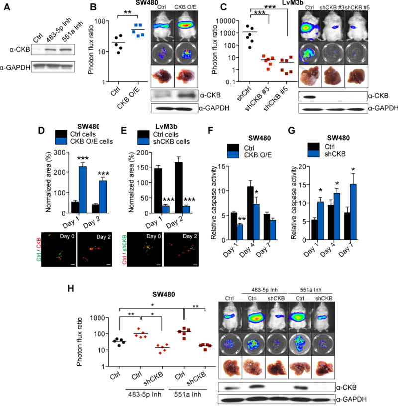
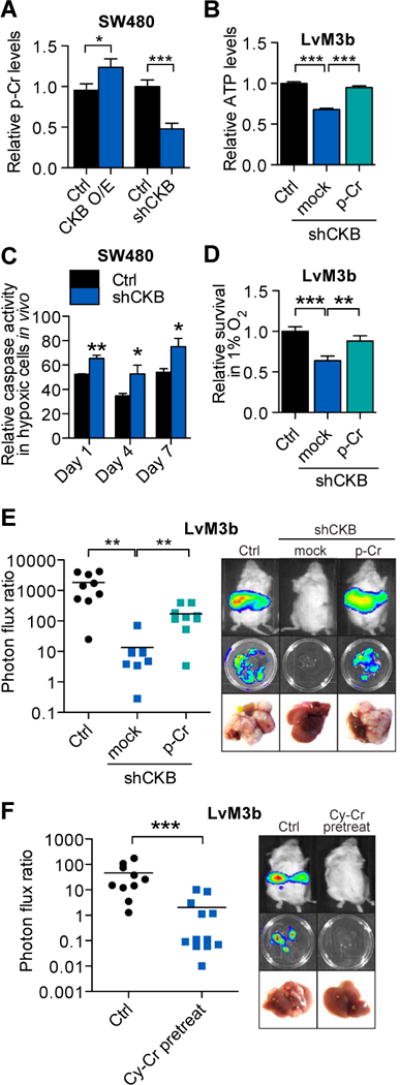
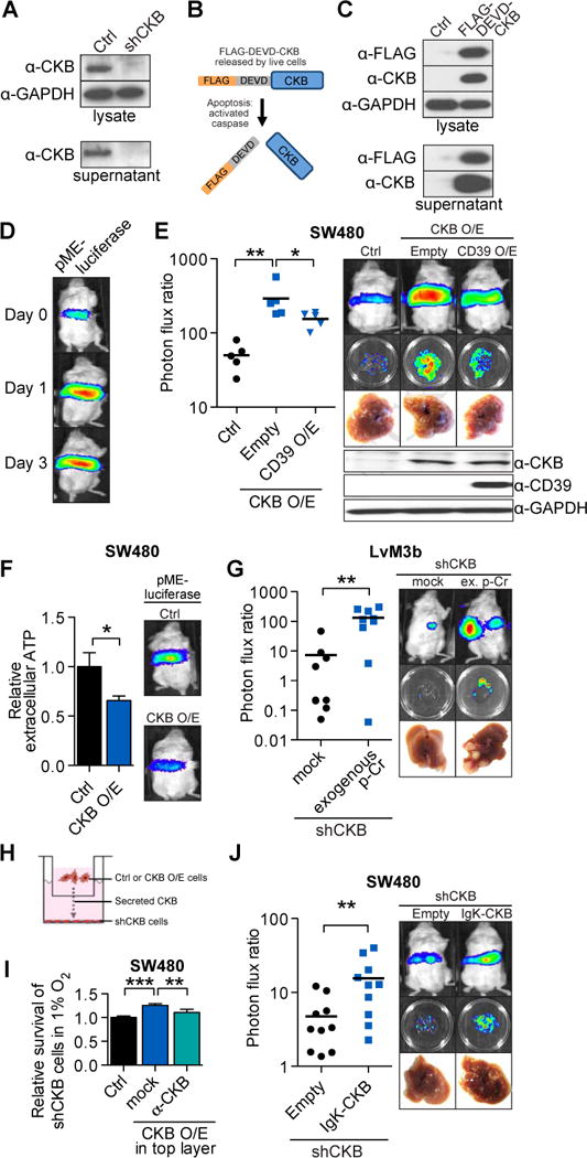
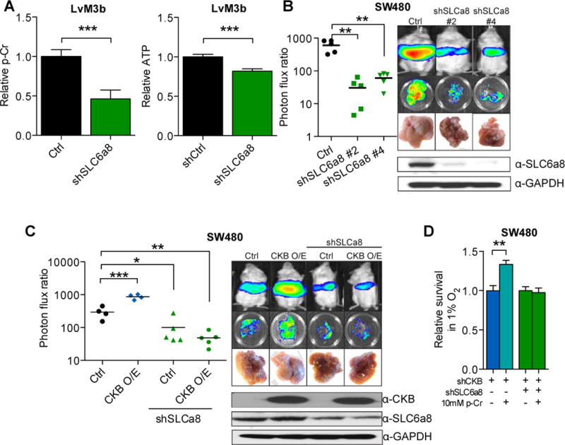
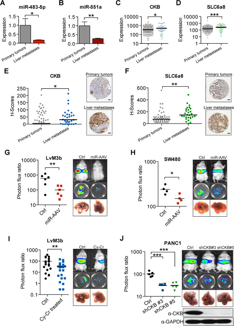
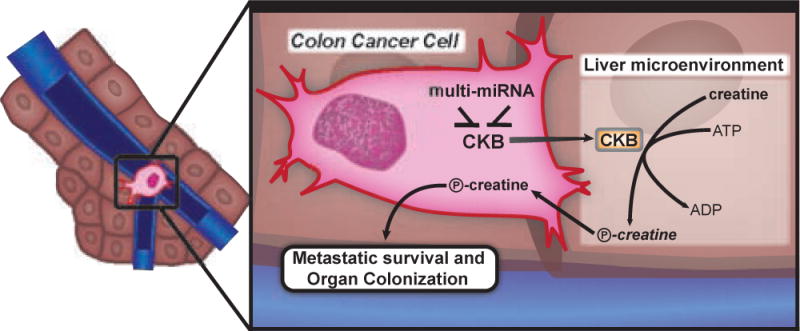
Colorectal cancer primarily metastasizes to the liver and globally kills over 600,000 people annually. By functionally screening 661 microRNAs (miRNAs) in parallel during liver colonization, we have identified miR-551a and miR-483 as robust endogenous suppressors of liver colonization and metastasis. These miRNAs convergently target creatine kinase, brain-type (CKB), which phosphorylates the metabolite creatine, to generate phosphocreatine. CKB is released into the extracellular space by metastatic cells encountering hepatic hypoxia and catalyzes production of phosphocreatine, which is imported through the SLC6A8 transporter and used to generate ATP—fueling metastatic survival. Combinatorial therapeutic viral delivery of miR-551a and miR-483-5p through single-dose adeno-associated viral (AAV) delivery significantly suppressed colon cancer metastasis, as did CKB inhibition with a small-molecule inhibitor. Importantly, human liver metastases express higher CKB and SLC6A8 levels and reduced miR-551a/miR-483 levels relative to primary tumors. We identify the extracellular space as an important compartment for malignant energetic catalysis and therapeutic targeting.


Figures

Figure 1
Figure 1. miR-483-5p and miR-551a are endogenous miRNAs that suppress liver metastasis
A, Bioluminescence plot of liver colonization by 5 × 10⁵ LS-Parental, LvM3a and LvM3b cells after direct intrahepatic injection (n>5). Mice were imaged at day 21 after injection and livers extracted for ex vivo imaging and gross morphological examination. Photon flux ratio is the ratio of bioluminescence signal at day 21 normalized to signal on day 0. B, Schematic for the identification of miR-483-5p and miR-551a as suppressors of metastasis. C, Liver metastasis of mice injected with 5 × 10⁵ LvM3b cells over-expressing either a control hairpin, miR-483-5p or miR-551a (n>5). D, Liver metastasis in mice injected with 5 × 10⁵ SW480 cells, whose endogenous miR-483-5p or miR-551a was inhibited (n>5). E, F, Organotypic slice culture imaging of SW480 cells (n=8) whose endogenous miR-483-5p (E) or miR-551a (F) were inhibited by LNAs. 5 × 10⁵ cells were labeled with cell-tracker green (control LNA) or cell-tracker red (miRNA specific LNA) and introduced into the livers prior to slice culture. Dye-swap experiments were performed to compensate for dye bias. Representative images at day 0 and day 3 are shown. Total area of each cell population at indicated time points are measured and normalized to start of experiment. Scale bar is equal to 50um. G, Bioluminescent metastatic signal from mice (n=5) injected with 5 × 10⁵ SW480 cells whose endogenous miR-483-5p or miR-551a activities were inhibited. Images and measurements were taken 24hr after tumor cells inoculation. H, Relative in vivo caspase activity of SW480 cells whose endogenous miR-483-5p or miR-551a was inhibited (n=3). Caspase activity was monitored using a caspase-3/7 activated DEVD-luciferin and normalized with bioluminescent signal from regular luciferin. Error bars, s.e.m; all P values are based on one-sided Student’s t-tests, or where appropriate, Mann-Whitney test for non-Gaussian distribution. *p<0.05; **p<0.01; ***p<0.001. See also Figure S1.
Figure 2
Figure 2. miR-483-5p and miR-551a suppress colorectal cancer cells survival and metastasis in the liver through regulation of CKB
A, Expression of CKB in SW480 cells whose endogenous miR-483-5p or miR-551a was inhibited with LNAs. B, Liver metastasis in mice injected intrasplenically with 5 × 10⁵ control SW480 cells and CKB over-expressing SW480 cells (n=5). Mice were euthanized at 28 days after injection. C, Liver metastasis in mice injected intrasplenically with 5 × 10⁵ LvM3b expressing a control hairpin or two independent shRNA hairpins targeting CKB (n=6). Mice were euthanized 21 days after injection. D, Survival of control SW480 and CKB over-expressing SW480 cells in organotypic liver slices (n=8). E, Survival of LvM3b cells expressing a control hairpin or hairpin targeting CKB in organotypic slice cultures (n=8). Representative images at day 0 and day 2 are shown. Scale bar represent 50um. F, Relative in vivo caspase activity of control or CKB over-expressing SW480 cells Caspase activity was measured on day 1, 4 and 7 after injection (n=3). G, Relative in vivo caspase-3 activity of SW480 cells expressing a control shRNA or shRNA targeting CKB. Caspase activity was measured on day 1, 4 and 7 after injection (n=3). H, Liver metastasis in mice injected with 5 × 10⁵ SW480 cells whose endogenous miR-483-5p or miR-551a was inhibited by LNA, with and without CKB knockdown. Error bars, s.e.m; all P values are based on one-sided Student’s t-tests, or where appropriate, Mann-Whitney test for non-Gaussian distribution. *p<0.05; **p<0.01; ***p<0.001. See also Figure S2.
Figure 3
Figure 3. CKB modulates colorectal cancer cell survival during acute intrahepatic hypoxia through modulation of the phosphocreatine/ATP shuttle
A, Relative intracellular phosphocreatine levels in SW480 cells over-expressing CKB or depleted for CKB (n=5). B, Relative intracellular ATP levels in LvM3b cells depleted for CKB, with and without exogenous 10um phosphocreatine supplementation (n=5). C, In vivo caspase activity of control and CKB knockdown SW480 cells experiencing hypoxia within the livers of mice (n=3). D, Survival of colorectal cancer cells in hypoxia in vitro with and without CKB knockdown, and 10um phosphocreatine supplementation (n=3). E, Liver metastasis by CKB depleted LvM3b cells pre-incubated overnight with 10um phosphocreatine. 5 × 10⁵ cells were then inoculated into the liver of mice through intrasplenic injection. F, Liver metastasis in mice injected with 5 × 10⁵ LvM3b cells with and without pre-treatment with 10mM cyclocreatine for 48hrs. Error bars, s.e.m; all p values are based on one-sided Student’s t-tests, or where appropriate, Mann-Whitney test for non-Gaussian distribution. *p<0.05; **p<0.01; ***p<0.001. See also Figure S3.
Figure 4
Figure 4. CKB is secreted by colorectal cancer cells and promotes malignant conversion of extracellular ATP and liver creatine to phosphocreatine to enhance metastasis
A, Extracellular and intracellular CKB protein levels in control and LvM3b cells depleted of CKB through RNAi. B, FLAG-tagged CKB with a caspase 3/7 recognition site linker. The FLAG-DEVD-CKB has a FLAG-tag linked to the N-terminal of CKB by a linker containing a caspase 3/7 recognition motif (DEVD-amino sequence). Caspase activation in apoptotic cells will result in cleavage of linker and release of FLAG-tag. C, Western-blot of FLAG-DEVD-CKB over-expressing cells demonstrate release of CKB by live cells into the extracellular space. D, Bioluminescent imaging of immunodeficient mice injected with SW480 cells expressing pME-Luc for detection of extracellular ATP (n=5). E, Liver metastasis by 5 × 10⁵ SW480 cells over-expressing CKB with concomitant over-expression of CD39. F, Relative extracellular ATP levels in CKB over-expressing cells. Control and CKB over-expressing pME-Luc SW480 cells were injected into mice (n=5). G, Liver metastasis by CKB-depleted LvM3b cells in mice implanted with an osmotic pump releasing phosphocreatine into the portal circulation. H, Scheme for co-culture experiment. 5 × 10⁴ CKB-knockdown cells were cultured on the bottom of 24-well plates, while control or CKB-over-expressing cells were plated onto boyden chambers above CKB-knockdown cells with pores for exchange of metabolites and proteins. Cells were counted after 4 days in hypoxia. I, Relative survival of CKB-knockdown cells in 1% oxygen when co-cultured with control, CKB-over-expressing cells or with CKB-over-expressing cells in the presence of a neutralizing antibody (n=4). J, Liver metastasis by endogenous CKB-knockdown SW480 cells over-expressing a secreted form of CKB. Error bars, s.e.m; all P values are based on one-sided Student’s t-tests, or where appropriate, Mann-Whitney test for non-Gaussian distribution. *p<0.05; **p<0.01; ***p<0.001. See also Figure S4.
Figure 5
Figure 5. SLC6a8 modulates CKB-mediated colon cancer metastasis through modulation of intracellular phosphocreatine levels
A, Relative intracellular phosphocreatine and ATP levels in LvM3b cells expressing a control shRNA or shRNA targeting SLC6a8 (n=4). B, Liver metastasis by 5 × 10⁵ LvM3b cells expressing two independent short hairpins targeting SLC6a8 (n=5). C, Liver metastasis by SW480 cells over-expressing CKB with and without SLC6a8 depletion (n>4). D, In vitro survival of LvM3b cells depleted of CKB, SLC6a8 with and without phosphocreatine supplementation in hypoxia (n=3). Error bars, s.e.m; all P values are based on one-sided Student’s t-tests, or where appropriate, Mann-Whitney test for non-Gaussian distribution. *p<0.05; **p<0.001; ***p<0.0001. See also Figure S5.
Figure 6
Figure 6. miR-483-5p, miR-551a, CKB and SLC6a8 are clinically relevant in independent cohorts of patients and can be therapeutically targeted
A, B, miR-483-5p and miR-551a levels in 36 primary colorectal adenocarcinomas and 30 liver metastases were quantified by quantitative real-time PCR. C, D, CKB and SLC6a8 expression from a public microarray dataset (GSE41258) comparing primary tumors and liver metastases (N=233). E, CKB expression in primary tumors compared to liver metastases examined through immunohistochemical staining of a tissue microarray (N=92). Scale bar represents 50um. F, SLC6a8 expression in primary tumors compared to liver metastases examined through immunohistochemical staining of above mentioned tissue microarray (N=88). Scale bar represents 50um. G, Liver metastasis in mice injected with 5 × 10⁵ LvM3b cells and treated with a single dose of AAV doubly expressing miR-483-5p and miR-551a one day after injection cells (n=6). H, Liver metastasis in mice injected with 5 × 10⁵ SW480 cells and treated with a single dose of AAV doubly expressing miR-483-5p and miR-551a one day after injection cells (n=4). I, Liver metastasis in mice injected with 5 × 10⁵ LvM3b cells and treated with cyclocreatine daily for two weeks (n>15). J, Liver metastasis by pancreatic cancer cells, PANC1, with knockdown of CKB with two independent shRNA hairpins (n=5). Error bars, s.e.m; all P values are based on one-sided Student’s t-tests, or where appropriate, Mann-Whitney test for non-Gaussian distribution. *p<0.05; **p<0.01; ***p<0.001.
Figure 7
Figure 7. Model for the miR-483-5p, miR-551a, CKB and SLC6a8 axis
Disseminated colon cancer cells arrive in the liver microenvironment through the hypoxemic portal circulation. Within the liver microenvironment, they experience hypoxic stress and ATP depletion. Cells that up-regulate CKB through loss of miRNAs, release CKB into the extracellular matrix where it converts available creatine and ATP into phosphocreatine that is then taken up by the cell to fuel metastatic survival and subsequent organ colonization. Colon cancer cells with higher levels of CKB also build up a larger pool of intracellular phosphocreatine that acts as a buffer against energetic stress.

Comment in

References

    1. Arteel GE, Thurman RG, Yates JM, Raleigh JA. Evidence that hypoxia markers detect oxygen gradients in liver: pimonidazole and retrograde perfusion of rat liver. Br J Cancer. 1995;72:889–895. - PMC - PubMed
    1. Bertout JA, Patel SA, Simon MC. The impact of O2 availability on human cancer. Nat Rev Cancer. 2008;8:967–975. - PMC - PubMed
    1. Bruns CJ, Harbison MT, Kuniyasu H, Eue I, Fidler IJ. In vivo selection and characterization of metastatic variants from human pancreatic adenocarcinoma by using orthotopic implantation in nude mice. Neoplasia. 1999;1:50–62. - PMC - PubMed
    1. Brustovetsky N, Brustovetsky T, Dubinsky JM. On the mechanisms of neuroprotection by creatine and phosphocreatine. J Neurochem. 2001;76:425–434. - PubMed
    1. Cairns RA, Harris IS, Mak TW. Regulation of cancer cell metabolism. Nat Rev Cancer. 2011;11:85–95. - PubMed

Publication types

MeSH terms

Associated data