. 2015 Feb 19;57(4):662-673.
doi: 10.1016/j.molcel.2014.12.023. Epub 2015 Jan 15.

WT1 recruits TET2 to regulate its target gene expression and suppress leukemia cell proliferation

Affiliations

WT1 recruits TET2 to regulate its target gene expression and suppress leukemia cell proliferation

Yiping Wang et al. Mol Cell. .

Abstract

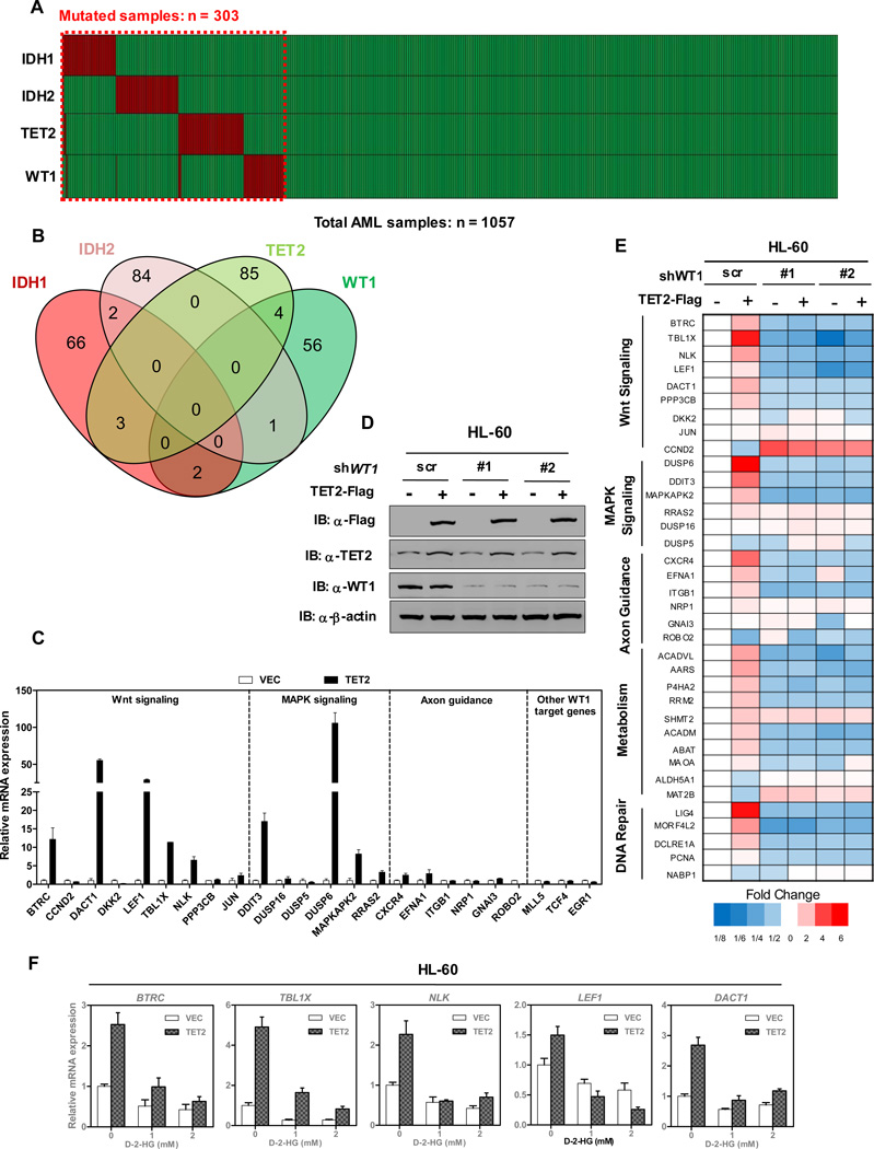
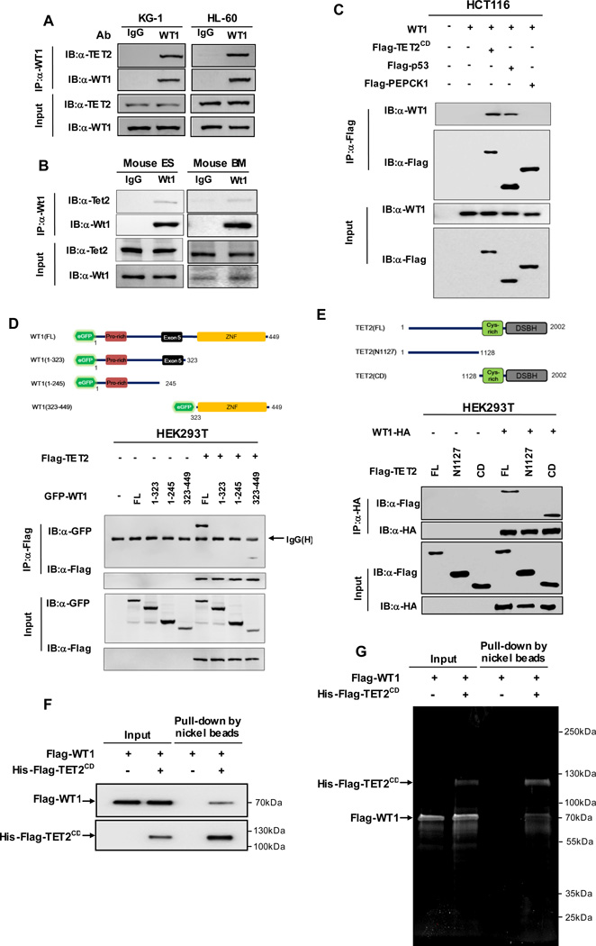
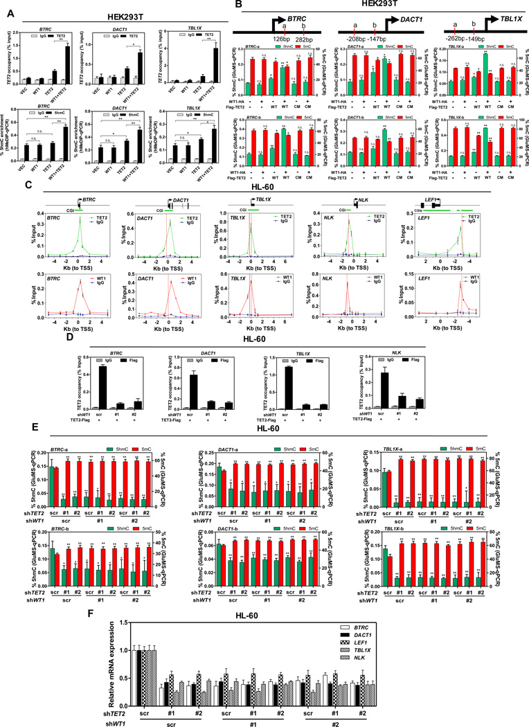
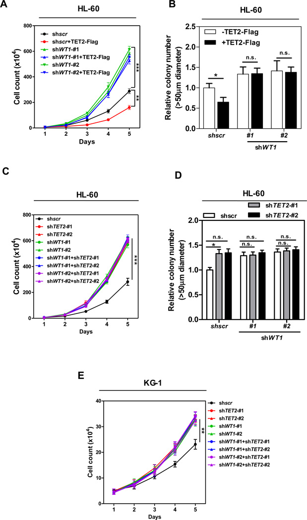
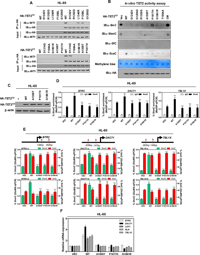
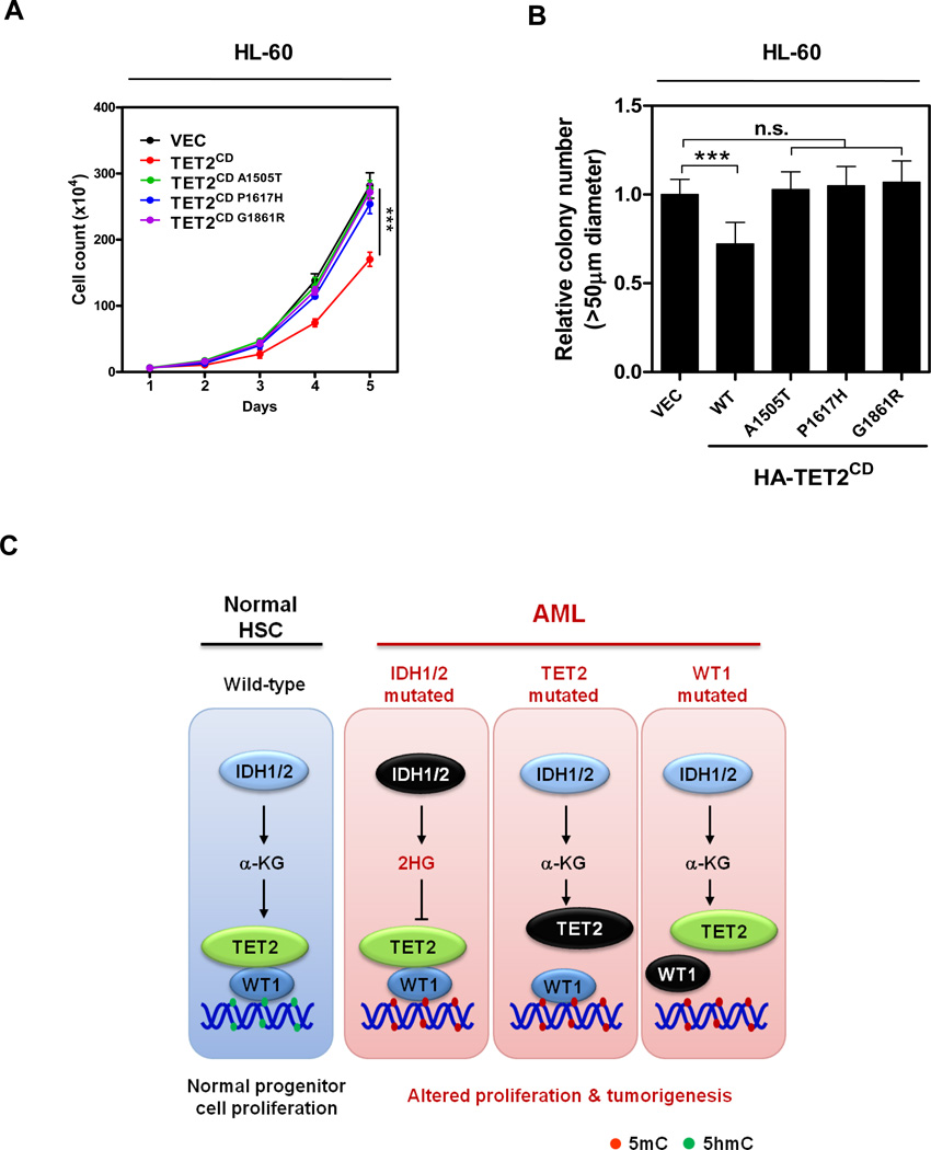
The TET2 DNA dioxygenase regulates cell identity and suppresses tumorigenesis by modulating DNA methylation and expression of a large number of genes. How TET2, like most other chromatin-modifying enzymes, is recruited to specific genomic sites is unknown. Here we report that WT1, a sequence-specific transcription factor, is mutated in a mutually exclusive manner with TET2, IDH1, and IDH2 in acute myeloid leukemia (AML). WT1 physically interacts with and recruits TET2 to its target genes to activate their expression. The interaction between WT1 and TET2 is disrupted by multiple AML-derived TET2 mutations. TET2 suppresses leukemia cell proliferation and colony formation in a manner dependent on WT1. These results provide a mechanism for targeting TET2 to a specific DNA sequence in the genome. Our results also provide an explanation for the mutual exclusivity of WT1 and TET2 mutations in AML, and suggest an IDH1/2-TET2-WT1 pathway in suppressing AML.


Figures

Figure 1
Figure 1. TET2 activates WT1 target genes
(A) Somatic variants in IDH1/2, TET2 and WT1 were identified in total 1057 AML cases, in which 303 cases carried at least one mutation. Data were collected from six different studies (2013; Fernandez-Mercado et al., 2012; Liang et al., 2013; Patel et al., 2012; Rocquain et al., 2010; Welch et al., 2012) (B) Overlap of IDH1/2, TET2 and WT1 mutations in 303 mutated cases of AML. (C) Flag-tagged full-length TET2 was overexpressed in HEK293T cells, and the mRNA expression of indicated WT1-target genes was determined by qRT-PCR. (D) Overexpression of TET2 in stable HL-60 cells with or without WT1 knockdown. HL-60 cells were transduced with retrovirus expressing different shRNAs against WT1 and retrovirus expressing Flag-tagged full-length TET2. The expression of WT1 and TET2 proteins was determined by western blot. (E) Stable HL-60 cells were generated as described in (D), and the mRNA expression of WT1 target genes was determined by qRT-PCR. (F) Stable HL-60 cells overexpressing TET2-Flag or empty vector control were treated with the indicated concentrations of cell-permeable D-2-HG for 12 hrs. mRNA expression of WT1-target genes was determined by qRT-PCR. Shown are average values of triplicated results with standard deviation (S.D.). See also Figure S1.
Figure 2
Figure 2. TET2 directly binds to WT1
(A) Endogenous WT1 protein was immunoprecipitated from two human AML cell lines (i.e. KG-1 and HL-60), following by western blot to detect TET2. Normal rabbit IgG was used as a negative control. (B) Endogenous Wt1 protein was immunoprecipitated from mouse ES cells or bone marrow (BM) cells, following by western blot to detect Tet2. Normal rabbit IgG was used as a negative control. (C) HCT116 cells were transiently transfected with plasmids expressing indicated genes. Protein-protein interaction was examined by IP-western using indicated antibodies. (D) Wild-type WT1 and its deletion mutants, as shown in the schematic illustration, were co-expressed with Flag-TET2 in HEK293T cells. Protein-protein interaction was examined by IP-western using indicated antibodies. (E) Wild-type TET2 and its deletion mutants, as shown in the schematic illustration, were co-expressed with WT1-HA in HEK293T cells. Protein-protein interaction was examined by IP-western using indicated antibodies. (F–G) Recombinant Flag-6xHis-TET2CD (200 ng) and Flag-WT1 (400 ng) proteins purified from baculovirus infected Sf9 cells were incubated together. Nickel beads were then added and bound proteins were eluted with imidazole and resolved by SDS-PAGE. WT1-TET2CD binding was examined by either western blot (F) or SYPRO Ruby staining (G). See also Figure S2.
Figure 3
Figure 3. TET2 is recruited by WT1 to its target genes
(A) Flag-TET2 was transiently expressed either singularly or with WT1-HA in HEK293T cells. The occupancy of Flag-TET2 at the promoter regions of WT1-target genes was determined by ChIP-qPCR (upper), and the 5hmC enrichment at the Flag-TET2 binding sites was determined by hMeDIP-qPCR (lower). Mouse IgG and rabbit IgG were included as negative controls for ChIP-qPCR and hMeDIP-qPCR, respectively. (B) Flag-tagged wild-type (WT) TET2 or its catalytic inactive mutant (CM) of TET2 was transiently overexpressed either singularly or with HA-WT1 in HEK293T cells. The site-specific 5hmC and 5mC levels were determined by using GluMS-qPCR. a, b indicate MspI/HapII recognition sites of each target gene. Arrow denotes promoter orientation. (C) Stable HL-60 cells with knockdown of endogenous TET2 and put-back of Flag-tagged TET2 were generated as described in Figure S3C. The occupancy of TET2-Flag and endogenous WT1 on the promoter regions of indicated WT1-target genes was determined by ChIP-qPCR. Mouse IgG and rabbit IgG were included as negative controls. Arrow denotes promoter orientation, CGI (green line) indicates CpG islands, red dash line indicates WT1 binding motif. (D) Stable HL-60 cells in (C) were transduced with retrovirus expressing different shRNAs against WT1. The occupancy of TET2-Flag on the promoter regions of indicated WT1-target genes was determined by ChIP-qPCR. Mouse IgG was included as negative control. (E) HL-60 cells were transduced with retrovirus expressing different shRNAs against WT1 and/or TET2 as described in Figure S3F. Site-specific levels of 5hmC and 5mC were determined by using GluMS-qPCR. (F) Stable HL-60 cells in (E) were examined for the mRNA expression of indicated WT1-target genes, as determined by qRT-PCR. Shown are average values of triplicated results with standard deviation (S.D.). * denotes P < 0.05 for the indicated comparison;** denotes P < 0.01 for the indicated comparison; n.s. = not significant. See also Figure S3.
Figure 4
Figure 4. TET2 inhibits leukemia cell proliferation in a WT1-dependent manner
(A–B) Cell proliferation (A) and colony formation (B) of stable HL-60 cells overexpressing full-length TET2 with or without WT1 knockdown were determined by cell counting and colony-forming assay, respectively. (C–D) Cell proliferation (C) and colony formation (D) of stable HL-60 cells with knockdown of WT1 and/or TET2 were determined by cell counting and colony-forming assay, respectively. (E) Cell proliferation of stable KG-1 cells with knockdown of WT1 and/or TET2 was determined by cell counting. Shown are average values of triplicated results with standard deviation (S.D.). *denotes the p < 0.05, **denotes the p < 0.01, and ***denotes the p < 0.001 for the indicated comparison. n.s.= not significant. See also Figure S4.
Figure 5
Figure 5. AML-derived mutations in TET2 disrupt WT1 binding
(A) HL-60 cells were transiently transfected with vectors encoding HA-tagged wild-type TET2CD, or AML-derived WT1-binding defective TET2 mutants as indicated, and the ectopically expressed TET2 proteins were immunoprecipitated, following by western blot to detect endogenous WT1. (B) In vitro TET2 catalytic activity assay. Genomic DNA were isolated from human monocyte-derived macrophage (MDM) cells, sonicated and incubated with immunopurified HA-tagged wild-type and mutants human TET2 at 37°C for 2 hrs. After termination of the reaction, a fraction of reaction mixture from each reaction was subjected to dot-blot assay using the antibodies specific for 5mC, 5hmC, 5fC and 5caC. See Experimental Procedures for more details. The amount of DNA in each reaction was examined by methylene blue staining. CM refers to a catalytic mutant of TET2 that harbors two mutations disrupting the binding with Fe2+ (C) HL-60 cells were transduced with lentiviral vectors encoding HA-tagged wild-type TET2CD, or AML-derived WT1-binding defective TET2CD mutants as indicated, and the expression of ectopic TET2CD proteins was determined by western blot. (D–F) Stable HL-60 cells were generated as described above in (C). The 5hmC enrichment at TET2 binding sites at the promoter regions of indicated WT1-target genes was determined by hMeDIP-qPCR (D), rabbit IgG was included as negative control. Moreover, the site-specific levels of 5hmC and 5mC were determined by using GluMS-qPCR (E), and the mRNA expression of indicated WT1-target genes was determined by qRT-PCR (F). Shown are average values of triplicated results with standard deviation (S.D.). *denotes the p < 0.05 and **denotes the p < 0.01 for the indicated comparison. n.s.= not significant. See also Figure S5.
Figure 6
Figure 6. AML-derived WT1 binding-defective TET2 mutants fail to suppress leukemia cell proliferation
(A–B) Cell proliferation (A) and colony formation (B) of stable HL-60 cells expressing wild-type or WT1 binding-defective mutants of HA-TET2CD were determined by cell counting and colony-forming assay, respectively. (C) A schematic illustration of the IDH1/2-TET2-WT1 pathway in AML suppression. See also Table S2.

Comment in

  • A new path to leukemia with WIT.
    Sardina JL, Graf T. Sardina JL, et al. Mol Cell. 2015 Feb 19;57(4):573-574. doi: 10.1016/j.molcel.2015.02.005. Mol Cell. 2015. PMID: 25699704

Similar articles

Cited by

References

    1. Cancer Genome Atlas Network. Genomic and epigenomic landscapes of adult de novo acute myeloid leukemia. N Engl J Med. 2013;368:2059–2074. - PMC - PubMed
    1. Chen Q, Chen Y, Bian C, Fujiki R, Yu X. TET2 promotes histone O-GlcNAcylation during gene transcription. Nature. 2013;493:561–564. - PMC - PubMed
    1. Chowdhury R, Yeoh KK, Tian YM, Hillringhaus L, Bagg EA, Rose NR, Leung IK, Li XS, Woon EC, Yang M, et al. The oncometabolite 2-hydroxyglutarate inhibits histone lysine demethylases. EMBO reports. 2011;12:463–469. - PMC - PubMed
    1. Costa Y, Ding J, Theunissen TW, Faiola F, Hore TA, Shliaha PV, Fidalgo M, Saunders A, Lawrence M, Dietmann S, et al. NANOG-dependent function of TET1 and TET2 in establishment of pluripotency. Nature. 2013;495:370–374. - PMC - PubMed
    1. Dawlaty MM, Breiling A, Le T, Raddatz G, Barrasa MI, Cheng AW, Gao Q, Powell BE, Li Z, Xu M, et al. Combined deficiency of Tet1 and Tet2 causes epigenetic abnormalities but is compatible with postnatal development. Dev Cell. 2013;24:310–323. - PMC - PubMed

Publication types

MeSH terms