. 2015 Jan 5:5:750.
doi: 10.3389/fpls.2014.00750. eCollection 2014.

Alternative translational initiation of ATP sulfurylase underlying dual localization of sulfate assimilation pathways in plastids and cytosol in Arabidopsis thaliana

Affiliations

Alternative translational initiation of ATP sulfurylase underlying dual localization of sulfate assimilation pathways in plastids and cytosol in Arabidopsis thaliana

Anne-Sophie Bohrer et al. Front Plant Sci. .

Abstract

Plants assimilate inorganic sulfate into sulfur-containing vital metabolites. ATP sulfurylase (ATPS) is the enzyme catalyzing the key entry step of the sulfate assimilation pathway in both plastids and cytosol in plants. Arabidopsis thaliana has four ATPS genes (ATPS1, -2, -3, and -4) encoding ATPS pre-proteins containing N-terminal transit peptide sequences for plastid targeting, however, the genetic identity of the cytosolic ATPS has remained unverified. Here we show that Arabidopsis ATPS2 dually encodes plastidic and cytosolic ATPS isoforms, differentiating their subcellular localizations by initiating translation at AUG(Met1) to produce plastid-targeted ATPS2 pre-proteins or at AUG(Met52) or AUG(Met58) within the transit peptide to have ATPS2 stay in cytosol. Translational initiation of ATPS2 at AUG(Met52) or AUG(Met58) was verified by expressing a tandem-fused synthetic gene, ATPS2 (5'UTR-His12) :Renilla luciferase:ATPS2 (Ile13-Val77) :firefly luciferase, under a single constitutively active CaMV 35S promoter in Arabidopsis protoplasts and examining the activities of two different luciferases translated in-frame with split N-terminal portions of ATPS2. Introducing missense mutations at AUG(Met52) and AUG(Met58) significantly reduced the firefly luciferase activity, while AUG(Met52) was a relatively preferred site for the alternative translational initiation. The activity of luciferase fusion protein starting at AUG(Met52) or AUG(Met58) was not modulated by changes in sulfate conditions. The dual localizations of ATPS2 in plastids and cytosol were further evidenced by expression of ATPS2-GFP fusion proteins in Arabidopsis protoplasts and transgenic lines, while they were also under control of tissue-specific ATPS2 promoter activity found predominantly in leaf epidermal cells, guard cells, vascular tissues and roots.

Keywords: ATP sulfurylase; Arabidopsis; alternative translational initiation; dual localization; sulfur metabolism.


Figures

Figure 1
Figure 1
Alignment of Arabidopsis ATP sulfurylase (ATPS) proteins. Arabidopsis thaliana ATPS complete protein sequences were aligned using Clustal W2. The predicted cleavage site of the transit peptide for plastid targeting is indicated. Identical residues in all four sequences are shaded and methionine residues in the transit peptides are boxed.
Figure 2
Figure 2
Alternative translational initiation sites of ATPS2 determined by expression of dual-luciferase-tagged fusion constructs in Arabidopsis protoplasts. (A) Schematic representation of p35S:ATPS2-dual-Luc used for expression of tandem fusion gene ATPS2(5′UTR-His12):Renilla luciferase:ATPS2(Ile13−Val77):firefly luciferase. Positions of annealing sites of primers (1F-4R) used for chimeric gene construction and stop codons (X) are indicated. (B) FLuc activity of protein extracts from Arabidopsis protoplasts transfected with either wild-type (WT) or mutated versions of p35:ATPS2-dual-Luc chimeric genes. Results are presented as FLuc/Rluc activity ratios in the same transfection. Values indicate mean ± SD for the results of 8 independent transfections. Values marked with different letters indicate statistically significant differences (ANOVA followed by Tukey's HSD post-hoc test; p < 0.05).
Figure 3
Figure 3
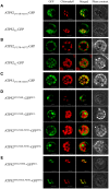
Subcellular localization of ATPS2-GFP fusion proteins in Arabidopsis protoplasts. (A) Chloroplast-cytosol dual-localization of ATPS2-GFP (ATPS2(5′UTR-Val77)-GFP and ATPS2FL-GFP). (B) Cytosolic localization of ATPS2-GFP (ATPS2(5′UTR-Val77)-GFP and ATPS2FL-GFP) in mesophyll protoplasts. (C) Chloroplastic localization of ATPS1-GFP (ATPS1(5′UTR-Val63)-GFP) in mesophyll protoplasts. (D) Cytosolic localization of ATPS2M1L/M4L-GFPM1L, ATPS2M1L/M4L/M52L-GFPM1L, and ATPS2M1L/M4L/M58L-GFPM1L. (E) Chloroplastic localization of ATPS2M52L/M58L-GFPM1L and ATPS2M4L/M52L/M58L-GFPM1L. Fluorescence was detected using a confocal laser-scanning microscope. GFP fluorescence (green), chlorophyll fluorescence (red), merged images (green and red) and bright-field phase contrast images are shown. Scale bars = 10 μm.
Figure 4
Figure 4
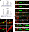
Tissue and subcellular localization of ATPS2-GFP fusion proteins in Arabidopsis transgenic plants. (A) Detection of ATPS2-GFP fusion proteins in each transgenic line by western blotting using anti-GFP antibody. (B) GFP fluorescence (green) observed in transverse sections of leaves from the transgenic lines expressing ATPS1pro:ATPS1:GFP, ATPS2pro:ATPS2:GFP and mutated versions (ATPS2M1I/M4I-GFP and ATPS2M52I/M58I-GFP). Red indicates chlorophyll autofluorescence. Guard cells (gc), mesophyll cells (m) and vascular tissues (vasc) are indicated. Scale bars = 100 μm. (C–H) Subcellular localizations of GFP fluorescence (green) in leaves (L), roots (R), guard cells (Gc), and mesophyll cells (M). Red indicates chlorophyll autofluorescence. Yellow or orange indicates overlap between green and red signals. Scales bars = 20 μm (for leaves), 100 μm (for roots), and 10 μm (for guard cells and mesophyll cells).

References

    1. Andréasson E., Bolt Jørgensen L., Höglund A.-S., Rask L., Meijer J. (2001). Different myrosinase and idioblast distribution in Arabidopsis and Brassica napus. Plant Physiol. 127, 1750–1763. 10.1104/pp.010334 - DOI - PMC - PubMed
    1. Aubry S., Smith-Unna R. D., Boursnell C. M., Kopriva S., Hibberd J. M. (2014). Transcript residency on ribosomes reveals a key role for the Arabidopsis thaliana bundle sheath in sulfur and glucosinolate metabolism. Plant J. 78, 659–673. 10.1111/tpj.12502 - DOI - PubMed
    1. Bradford M. M. (1976). A rapid and sensitive method for the quantitation of microgram quantities of protein utilizing the principle of protein-dye binding. Anal. Biochem. 72, 248–254. 10.1016/0003-2697(76)90527-3 - DOI - PubMed
    1. Chiu W., Niwa Y., Zeng W., Hirano T., Kobayashi H., Sheen J. (1996). Engineered GFP as a vital reporter in plants. Curr. Biol. 6, 325–330. 10.1016/S0960-9822(02)00483-9 - DOI - PubMed
    1. Clough S. J., Bent A. F. (1998). Floral dip: a simplified method for Agrobacterium-mediated transformation of Arabidopsis thaliana. Plant J. 16, 735–743. 10.1046/j.1365-313x.1998.00343.x - DOI - PubMed

LinkOut - more resources