Review
. 2015 Jan 5:4:341.
doi: 10.3389/fonc.2014.00341. eCollection 2014.

Notching on Cancer's Door: Notch Signaling in Brain Tumors

Affiliations
Review

Notching on Cancer's Door: Notch Signaling in Brain Tumors

Marcin Teodorczyk et al. Front Oncol. .

Abstract

Notch receptors play an essential role in the regulation of central cellular processes during embryonic and postnatal development. The mammalian genome encodes for four Notch paralogs (Notch 1-4), which are activated by three Delta-like (Dll1/3/4) and two Serrate-like (Jagged1/2) ligands. Further, non-canonical Notch ligands such as epidermal growth factor like protein 7 (EGFL7) have been identified and serve mostly as antagonists of Notch signaling. The Notch pathway prevents neuronal differentiation in the central nervous system by driving neural stem cell maintenance and commitment of neural progenitor cells into the glial lineage. Notch is therefore often implicated in the development of brain tumors, as tumor cells share various characteristics with neural stem and progenitor cells. Notch receptors are overexpressed in gliomas and their oncogenicity has been confirmed by gain- and loss-of-function studies in vitro and in vivo. To this end, special attention is paid to the impact of Notch signaling on stem-like brain tumor-propagating cells as these cells contribute to growth, survival, invasion, and recurrence of brain tumors. Based on the outcome of ongoing studies in vivo, Notch-directed therapies such as γ-secretase inhibitors and blocking antibodies have entered and completed various clinical trials. This review summarizes the current knowledge on Notch signaling in brain tumor formation and therapy.

Keywords: Notch signaling; brain tumor therapy; clinical trials; glioma; medulloblastoma; stem-like brain tumor-propagating cells.


Figures
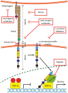
Figure 1
Figure 1
Canonical Notch signaling with points of intervention of current therapies. The interaction between Delta/Jagged-type ligands and Notch receptors leads to S2 cleavage on the extracellular site by “a disintegrin and metalloprotease” 10 (ADAM10) or ADAM17, which is followed by S3 cleavage by the γ-secretase–presenilin complex. The S3 cleavage gives rise to an intracellular Notch fragment (NICD) that translocates into the nucleus, where NICD binds to a protein complex containing recombination signal-binding protein Jκ (RBP-Jκ). This mediates the conversion of RBP-Jκ from a repressor to a transcriptional activator and is followed by the recruitment of the co-activator mastermind-like 1 (MAML1). These events lead to the de-repression of transcription of hairy/enhancer of split (Hes) and Hey. Several stages of the Notch signaling pathway are prone to pharmacological intervention and are labeled in the figure. Gamma-secretase inhibitors and blocking antibodies are already in clinical trials and decoys have been tested in animal models. Peptide inhibitors represent potential future treatment modalities. NECD, Notch extracellular domain; NTM, Notch transmembrane domain.
Figure 2
Figure 2
Notch signaling modules relevant for brain tumors. Notch signaling has been shown to be modulated on multiple levels in glioma cells and is linked upstream and downstream to other tumorigenic pathways. Its expression is induced by Ras and Akt, while Notch itself induces expression of epidermal growth factor (EGFR) via p53 (indirectly; hence dashed arrow) and pro-migratory glycoprotein tenascin C (TNC). Hypoxia-inducible factor 1α (HIF-1α) and HIF-2α compete for NICD binding. The Notch inhibitor HIF-2α is displaced by HIF-1α under hypoxic conditions. Further, several proteins modulate the Notch pathway at the level of NICD. Examples are Numb, which promotes Notch degradation via ubiquitin ligases such as FBW7 or Itch, or Numb4d7 and ligand of Notch protein X (LNX), which stimulate Notch signaling.

Similar articles

Cited by

References

    1. Louis DN, Ohgaki H, Wiestler OD, Cavenee WK, Burger PC, Jouvet A, et al. The 2007 WHO classification of tumours of the central nervous system. Acta Neuropathol (2007) 114(2):97–109.10.1007/s00401-007-0278-6 - DOI - PMC - PubMed
    1. Wen PY, Kesari S. Malignant gliomas in adults. N Engl J Med (2008) 359(5):492–50710.1056/NEJMra0708126 - DOI - PubMed
    1. Bralten LB, French PJ. Genetic alterations in glioma. Cancers (2011) 3(1):1129–4010.3390/cancers3011129 - DOI - PMC - PubMed
    1. Yoshida J. Molecular neurosurgery using gene therapy to treat malignant glioma. Nagoya J Med Sci (1996) 59(3–4):97–105. - PubMed
    1. Gajjar A, Chintagumpala M, Ashley D, Kellie S, Kun LE, Merchant TE, et al. Risk-adapted craniospinal radiotherapy followed by high-dose chemotherapy and stem-cell rescue in children with newly diagnosed medulloblastoma (St Jude Medulloblastoma-96): long-term results from a prospective, multicentre trial. Lancet Oncol (2006) 7(10):813–20.10.1016/S1470-2045(06)70867-1 - DOI - PubMed

LinkOut - more resources