Review
. 2015 Mar 5;370(1663):20140072.
doi: 10.1098/rstb.2014.0072.

A review of inter- and intraspecific variation in the eutherian placenta

Affiliations
Review

A review of inter- and intraspecific variation in the eutherian placenta

William E Gundling Jr et al. Philos Trans R Soc Lond B Biol Sci. .

Abstract

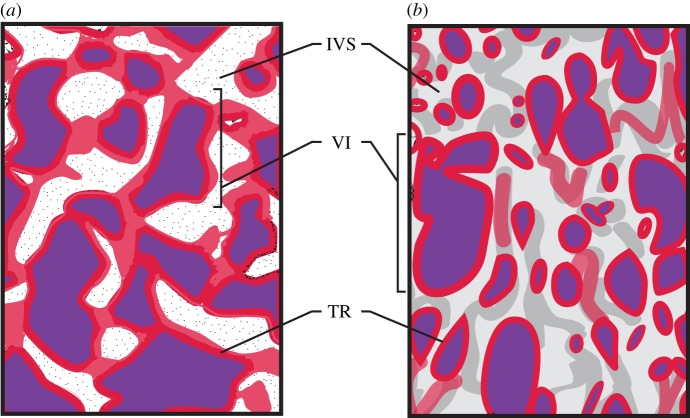
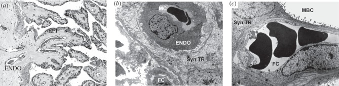
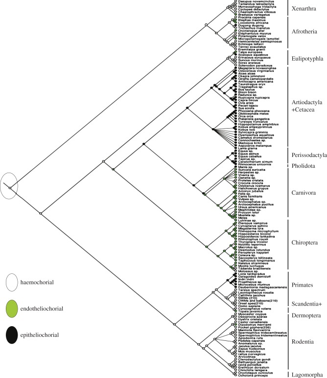
The placenta is one of the most morphologically variable mammalian organs. Four major characteristics are typically discussed when comparing the placentas of different eutherian species: placental shape, maternal-fetal interdigitation, intimacy of the maternal-fetal interface and the pattern of maternal-fetal blood flow. Here, we describe the evolution of three of these features as well as other key aspects of eutherian placentation. In addition to interspecific anatomical variation, there is also variation in placental anatomy and function within a single species. Much of this intraspecific variation occurs in response to different environmental conditions such as altitude and poor maternal nutrition. Examinations of variation in the placenta from both intra- and interspecies perspectives elucidate different aspects of placental function and dysfunction at the maternal-fetal interface. Comparisons within species identify candidate mechanisms that are activated in response to environmental stressors ultimately contributing to the aetiology of obstetric syndromes such as pre-eclampsia. Comparisons above the species level identify the evolutionary lineages on which the potential for the development of obstetric syndromes emerged.

Keywords: adaptation; placenta; variation.


Figures

Figure 1.
Figure 1.
Placental interdigitation across primates. Cross-sectional view of two types of maternal–fetal interdigitation found in primates: trabecular (a) and villous (b). The trabecular interdigitation pattern is characterized as branching globular folds that terminate in villi and this is seen in tarsiers and platyrrhines. The fetal trophoblast cells (red) surround the fetal villi (purple), which contain fetal blood (purple) separating the fetal blood from the maternal blood located in the intervillous space. The villous interdigitation pattern is found in catarrhines and strepsirrhines, and this is characterized by a branching villi pattern [4]. TR, trophoblast cells; VI, villi; IVS, maternal intervillous space. Adapted from [18]. (Online version in colour.)
Figure 2.
Figure 2.
Three types for interface between the mother and the fetus. Histological examples of the three different levels maternal–fetal interface seen across the eutherians. (a) Section of placental tissue from a giraffe which has an epitheliochorial interface where the mother and the fetus are separated by layer of uterine epithelial cells (EPI) and maternal endothelial cells (ENDO). (b) An example of an endotheliochorial placenta of a three toed sloth originally from [12]. This type of placenta has one layer of maternal endothelial cells (ENDO) separating maternal blood from the trophoblast. (c) An example of a haemochorial placenta of a degu, a rodent from [20]. The haemochorial placenta is the most invasive maternal–fetal interface with fetal trophoblast in direct contact with maternal blood. ENDO, maternal endothelial cells; EPI, maternal epithelial cells; Syn TR, syncytiotrophoblast; FC, fetal capillary; MBC, maternal blood channel. With permissions from the publishers and authors.
Figure 3.
Figure 3.
Evolution of the maternal–fetal interface. Maximum-parsimony phylogenetic reconstruction of the maternal–fetal interface of 141 mammals through parsimony analysis showing the root placental mammal having a haemochorial placenta (white) with epitheliochorial (black) and endotheliochorial (green) as divergent states [,,–24]. Tree topology taken from [25]. (Online version in colour.)

References

    1. Lewis RM, Cleal JK, Hanson MA. 2012. Placenta, evolution and lifelong health. Placenta 33, S28–S32. (10.1016/j.placenta.2011.12.003) - DOI - PubMed
    1. Lager S, Powell TL. 2012. Regulation of nutrient transport across the placenta. J. Pregnancy 2012, 179827 (10.1155/2012/179827) - DOI - PMC - PubMed
    1. Wildman DE, Chen C, Erez O, Grossman LI, Goodman M, Romero R. 2006. Evolution of the mammalian placenta revealed by phylogenetic analysis. Proc. Natl Acad. Sci. USA 103, 3203–3208. (10.1073/pnas.0511344103) - DOI - PMC - PubMed
    1. Benirschke K, Kaufmann P. 2000. Pathology of the human placenta, 4th edn New York, NY: Springer.
    1. Zamudio S, Palmer SK, Dahms TE, Berman JC, Young DA, Moore LG. 1995. Alterations in uteroplacental blood flow precede hypertension in preeclampsia at high altitude. J. Appl. Physiol. 79, 15–22. - PubMed

Publication types