. 2015 May;135(5):1320-1328.
doi: 10.1038/jid.2015.6. Epub 2015 Jan 20.

Genetic and pharmacological analysis identifies a physiological role for the AHR in epidermal differentiation

Affiliations

Genetic and pharmacological analysis identifies a physiological role for the AHR in epidermal differentiation

Ellen H van den Bogaard et al. J Invest Dermatol. 2015 May.

Abstract

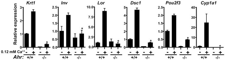
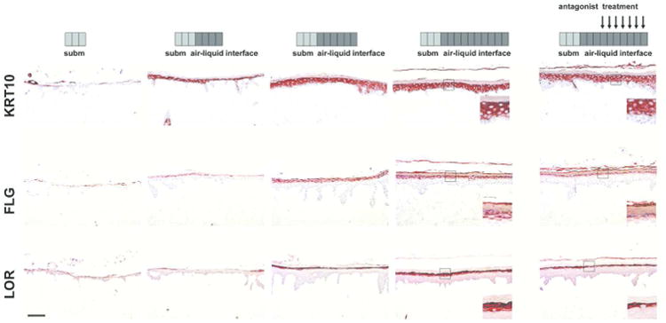
Stimulation of the aryl hydrocarbon receptor (AHR) by xenobiotics is known to affect epidermal differentiation and skin barrier formation. The physiological role of endogenous AHR signaling in keratinocyte differentiation is not known. We used murine and human skin models to address the hypothesis that AHR activation is required for normal keratinocyte differentiation. Using transcriptome analysis of Ahr(-/-) and Ahr(+/+) murine keratinocytes, we found significant enrichment of differentially expressed genes linked to epidermal differentiation. Primary Ahr(-/-) keratinocytes showed a significant reduction in terminal differentiation gene and protein expression, similar to Ahr(+/+) keratinocytes treated with AHR antagonists GNF351 and CH223191, or the selective AHR modulator (SAhRM) SGA360. In vitro keratinocyte differentiation led to increased AHR levels and subsequent nuclear translocation, followed by induced CYP1A1 gene expression. Monolayer cultured primary human keratinocytes treated with AHR antagonists also showed an impaired terminal differentiation program. Inactivation of AHR activity during human skin equivalent development severely impaired epidermal stratification, terminal differentiation protein expression, and stratum corneum formation. As disturbed epidermal differentiation is a main feature of many skin diseases, pharmacological agents targeting AHR signaling or future identification of endogenous keratinocyte-derived AHR ligands should be considered as potential new drugs in dermatology.


Conflict of interest statement

Conflict of Interest: The authors have no conflict of interest to declare.

Figures

Figure 1
Figure 1. Downregulation of differentiation gene expression in Ahr-/- mouse keratinocytes
(a) Expression of indicated genes during calcium induced differentiation of primary Ahr+/+ and Ahr-/- keratinocytes was determined by quantitative PCR from triplicate cultures, repeated twice. Expression was normalized to Gapdh. * significantly different from Ahr +/+ p<.05. Krt1, Keratin 1; Lor, Loricrin; Ivl, Involucrin; Dsc1, Desmocollin 1; Pou2f3, POU Class 2 Homeobox 3, Cyp1a1, Cytochrome P450 1A1.
Figure 2
Figure 2. AHR antagonists and selective modulators suppress epidermal differentiation in monolayer culture
(a) Effect of TCDD (10 nM); GNF351 (500 nM) and SGA360 (10 μM) on gene expression in proliferating (0.05 mM CaCl2) or differentiating (0.12 mM CaCl2) primary mouse keratinocytes (triplicate, repeated twice) * p<.05 compared to vehicle control (b) Effect of GNF351 (500 nM); CH223191 (CH, 5 μM) and indirubin (IR, 50 nM) on differentiation gene expression in primary human keratinocytes. (2 separate experiments, total of n=5 donors). (c) Immunoblot analysis showing effect of Ahr ablation, GNF351 or SGA360 on differentiation induced expression of keratin 10 and loricrin in primary mouse keratinocytes. (d) Immunoblot analysis showing effect of GNF351 and CH223191 (CH) on pro-filaggrin (FLG), involucrin (IVL) and loricrin (LOR) in monolayer cultured primary human keratinocytes.
Figure 3
Figure 3. Epidermal stratification defects and reduced stratum corneum thickness caused by AHR inactivation
Human skin equivalents (epidermis-only) were generated on plastic inert filters. At indicated time points (arrows) during skin equivalent development (each block represents one day of culture), AHR antagonists were added to the culture medium. All skin equivalents were harvested at day 10 of air-liquid interface culture. (a) Hematoxylin and Eosin staining of skin equivalents treated with GNF351 (500 nM) or CH223191 (CH) (5 μM). (b) Immunohistochemical staining of Keratin 10 (KRT10, early differentiation), filaggrin (FLG, terminal differentiation), involucrin (IVL, terminal differentiation) and Ki67 (proliferation) of skin equivalents treated with GNF351 as depicted in 2A. (n=2 keratinocyte donors). Scale bar = 100 μm.
Figure 4
Figure 4. Reduced terminal differentiation protein expression caused by AHR inactivation
Human skin equivalents were generated using de-epidermised dermis and expression of keratin 10 (KRT10), filaggrin (FLG) and loricrin (LOR) was followed in time by harvesting the skin equivalents directly after submerged culture, and after 4, 6 and 10 days of air-liquid interface culture (each block represents one day of culture). Treatment with GNF351 (500 nM, arrows) was initiated at day 6 and sustained until day 10 of air-liquid interface culture. Magnification inlays show epidermal differentiation protein expression affected by AHR inactivation. (n= 2 keratinocytes donors). Scale bar = 100 μm.
Figure 5
Figure 5. AHR antagonists suppress human keratinocyte proliferation
Monolayer cultures of human primary keratinocytes (n=3 keratinocyte donors, * p<0.05) were treated with AHR antagonists (GNF: GNF351, 500 nM; CH: CH223191, 5μM; SR1, 500 nM) for 48h during the proliferation stage of the culture. (a) Quantification of Ki67 positive cells and (b) total cell count of antagonist treated keratinocytes as compared to untreated cells.
Figure 6
Figure 6. Nuclear localization of AHR during epidermal differentiation
(a) Immunoblot of AHR and ARNT following induction of differentiation with elevated calcium medium. (b) Immunoblot of cytoplasmic and nuclear AHR in primary mouse keratinocytes induced to differentiate in presence of GNF351 or SGA360. (c) Immunoblot showing time course of nuclear and cytoplasmic AHR levels in response to AHR ligands in primary mouse keratinocytes cultured in proliferation media.

References

    1. Andersen B, Weinberg WC, Rennekampff O, et al. Functions of the POU domain genes Skn-1a/i and Tst-1/Oct-6/SCIP in epidermal differentiation. Genes Dev. 1997;11:1873–84. - PubMed
    1. Balato A, Lembo S, Mattii M, et al. IL-33 is secreted by psoriatic keratinocytes and induces pro-inflammatory cytokines via keratinocyte and mast cell activation. Exp Dermatol. 2012;21:892–4. - PubMed
    1. Boitano AE, Wang J, Romeo R, et al. Aryl hydrocarbon receptor antagonists promote the expansion of human hematopoietic stem cells. Science. 2010;329:1345–8. - PMC - PubMed
    1. Cameron GS, Baldwin JK, Jasheway DW, et al. Arachidonic acid metabolism varies with the state of differentiation in density gradient-separated mouse epidermal cells. J Invest Dermatol. 1990;94:292–6. - PubMed
    1. Carrier Y, Ma HL, Ramon HE, et al. Inter-regulation of Th17 cytokines and the IL-36 cytokines in vitro and in vivo: implications in psoriasis pathogenesis. J Invest Dermatol. 2011;131:2428–37. - PubMed

Publication types

MeSH terms