

Topical nepafenec in eyes with noncentral diabetic macular edema
- PMID: 25602634
- PMCID: PMC4408212
- DOI: 10.1097/IAE.0000000000000403
Topical nepafenec in eyes with noncentral diabetic macular edema
Abstract
Purpose: To evaluate the effect of a topical, nonsteroidal antiinflammatory drug, nepafenac 0.1%, in eyes with noncentral diabetic macular edema.
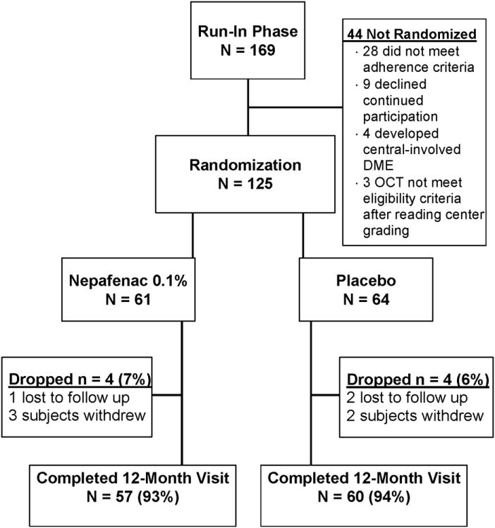
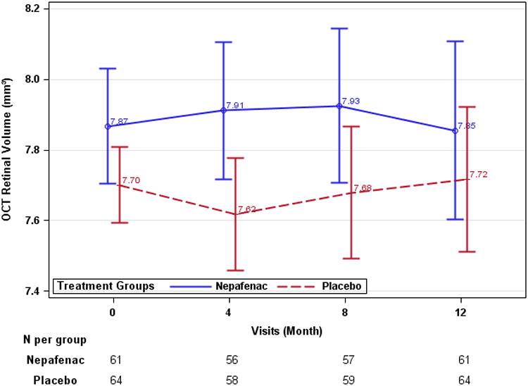
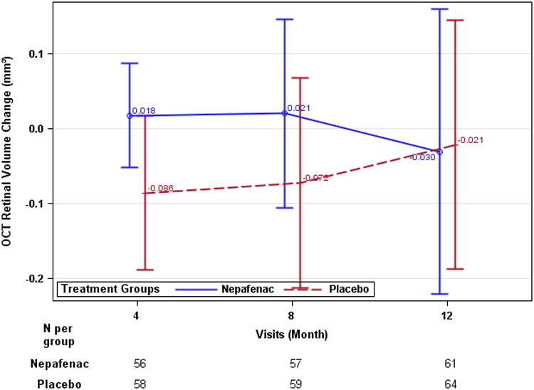
Methods: Multicenter, double-masked randomized trial. Individuals with good visual acuity and noncentral-involved diabetic macular edema were randomly assigned to nepafenac 0.1% (N = 61) or placebo (nepafenac vehicle, N = 64) 3 times a day for 12 months. The primary outcome was mean change in optical coherence tomography retinal volume at 12 months.
Results: Mean baseline retinal volume was 7.8 mm. At 12 months, in the nepafenac and placebo groups respectively, mean change in retinal volume was -0.03 mm and -0.02 mm (treatment group difference: -0.02, 95% confidence interval: -0.27 to 0.23, P = 0.89). Central-involved diabetic macular edema was present in 7 eyes (11%) and 9 eyes (14%) at the 12-month visit (P = 0.79), respectively. No differences in visual acuity outcomes were identified. One study participant developed a corneal melt after using nepafenac in the nonstudy eye, which had a history of severe dry eye. No additional safety concerns were evident.
Conclusion: In eyes with noncentral diabetic macular edema and good visual acuity, topical nepafenac 0.1% 3 times daily for 1 year likely does not have a meaningful effect on optical coherence tomography-measured retinal thickness.
Figures
References
-
- Bhagat N, Grigorian RA, Tutela A, et al. Diabetic macular edema: pathogenesis and treatment. Surv Ophthalmol. 2009;54:1–32. - PubMed
-
- National Diabetes Fact Sheet. Available at: http://www.cdc.gov/diabetes/pubs/factsheet11.htm.
-
- PKC-DMES Study Group. Effect of ruboxistaurin in patients with diabetic macular edema: thirty-month results of the randomized PKC-DMES clinical trial. Arch Ophthalmol. 2007;125:318–24. - PubMed
-
- Early Treatment Diabetic Retinopathy Study Research Group. Photocoagulation for diabetic macular edema Early Treatment Diabetic Retinopathy Study report number 1. Arch Ophthalmol. 1985;103:1796–806. - PubMed
Publication types
MeSH terms
Substances
Grants and funding
LinkOut - more resources
Full Text Sources
Medical
