. 2015 Jun 1;24(11):1342-51.
doi: 10.1089/scd.2014.0402. Epub 2015 Mar 3.

Catalase inhibits ionizing radiation-induced apoptosis in hematopoietic stem and progenitor cells

Affiliations

Catalase inhibits ionizing radiation-induced apoptosis in hematopoietic stem and progenitor cells

Xia Xiao et al. Stem Cells Dev. .

Abstract

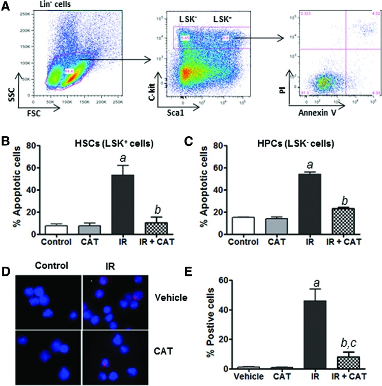
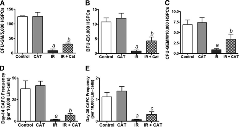
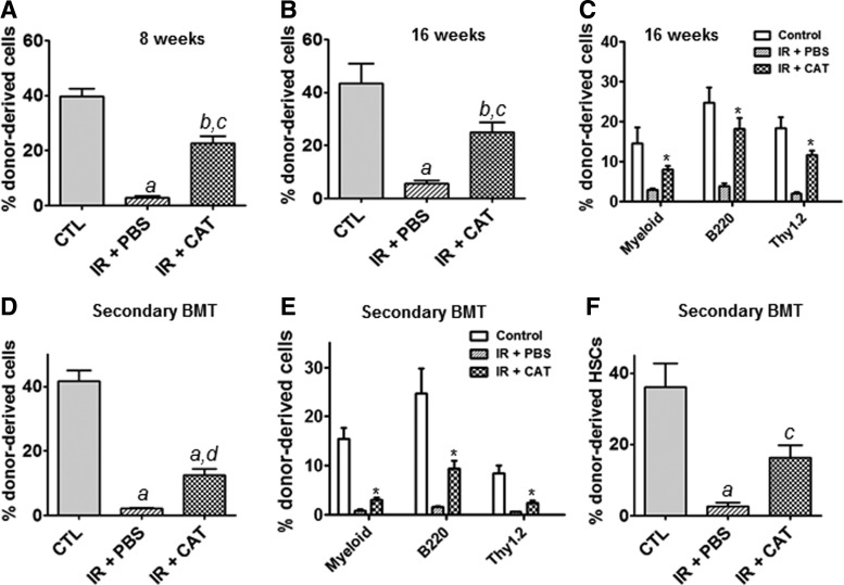
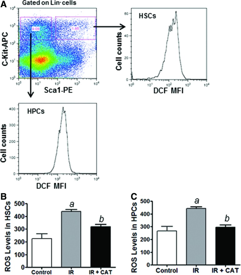
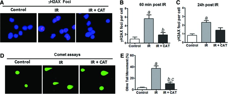
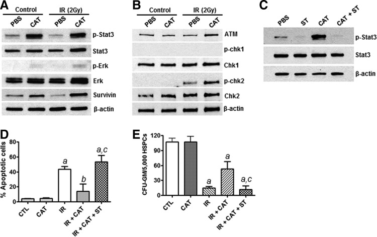
Hematologic toxicity is a major cause of mortality in radiation emergency scenarios and a primary side effect concern in patients undergoing chemo-radiotherapy. Therefore, there is a critical need for the development of novel and more effective approaches to manage this side effect. Catalase is a potent antioxidant enzyme that coverts hydrogen peroxide into hydrogen and water. In this study, we evaluated the efficacy of catalase as a protectant against ionizing radiation (IR)-induced toxicity in hematopoietic stem and progenitor cells (HSPCs). The results revealed that catalase treatment markedly inhibits IR-induced apoptosis in murine hematopoietic stem cells and hematopoietic progenitor cells. Subsequent colony-forming cell and cobble-stone area-forming cell assays showed that catalase-treated HSPCs can not only survive irradiation-induced apoptosis but also have higher clonogenic capacity, compared with vehicle-treated cells. Moreover, transplantation of catalase-treated irradiated HSPCs results in high levels of multi-lineage and long-term engraftments, whereas vehicle-treated irradiated HSPCs exhibit very limited hematopoiesis reconstituting capacity. Mechanistically, catalase treatment attenuates IR-induced DNA double-strand breaks and inhibits reactive oxygen species. Unexpectedly, we found that the radioprotective effect of catalase is associated with activation of the signal transducer and activator of transcription 3 (STAT3) signaling pathway and pharmacological inhibition of STAT3 abolishes the protective activity of catalase, suggesting that catalase may protect HSPCs against IR-induced toxicity via promoting STAT3 activation. Collectively, these results demonstrate a previously unrecognized mechanism by which catalase inhibits IR-induced DNA damage and apoptosis in HSPCs.


Figures

<b>FIG. 1.</b>
FIG. 1.
Catalase (CAT) inhibits IR-induced apoptosis in hematopoietic stem cells (HSCs) and hematopoietic progenitor cells (HPCs). (A) Schematic illustration of apoptosis analysis in Lin Sca1+ c-Kit+ (LSK+) HSCs and Lin Sca1+ c-Kit (LSK) HPCs. (B) The percentage of apoptotic cells in HSCs after IR and CAT treatment is presented as mean±SEM of three independent experiments. (C) The percentage of apoptotic cells in HPCs after IR and CAT treatment is presented as mean±SEM of three independent experiments. (D) Lin HSPCs were pretreated with CAT (100 U/mL) or PBS as vehicle control for 30 min before IR. Cleaved caspase-3 immunostaining was performed at 16 h after IR. Representative photomicrographs of activated caspase-3 immunofluorescent staining (red) and nucleic counterstaining with DAPI (blue) are shown. (E) The percentage of cells positively stained for cleaved caspase-3 in different groups of HSPCs is presented as mean±SEM of three independent assays. aP<0.001 versus PBS vehicle control; bP<0.01 versus IR; cP<0.05 versus PBS vehicle control. Color images available online at www.liebertpub.com/scd
<b>FIG. 2.</b>
FIG. 2.
CAT treatment protects the clonogenic function of irradiated HSPCs. (A–C) Colony-forming cell (CFC) assays were performed to determine the clonogenic capacity of HPCs to generate CFU-E, BFU-E, and CFU-GEMM as previously reported [3,6]. (D, E) CAFC assays were employed to examine the clonogenic functions of HPCs (day-14 CAFCs) and HSCs (day-35 CAFCs), respectively, as previously described [6,23]. Data are presented as mean±SEM of three independent experiments. aP<0.01 versus PBS vehicle control; bP<0.05 versus IR; cP<0.01 versus IR.
<b>FIG. 3.</b>
FIG. 3.
Transplantation of CAT-rescued irradiated HSPCs produces long-term engraftment in recipient mice. Competitive repopulation assays (CRA) were performed to determine the self-renewal and multi-lineage differentiation capacity of HSCs. (A) Short-term donor cell-derived engraftment in peripheral blood was determined at 8 weeks post transplantation using flow cytometric analyses. (B) Long-term donor cell-derived engraftment in peripheral blood was determined at 16 weeks after transplantation. (C) Shown is the percentage of donor cell-derived myeloid, B-, and T-lymphocytic lineage engraftment at 16 weeks post transplantation. Data are presented as mean±SEM (n=10 mice per group). (D) Donor cell-derived engraftment in peripheral blood was determined at 8 weeks after secondary bone marrow transplantation (BMT). (E) Shown is the percentage of donor cell-derived myeloid, B-, and T-lymphocytic lineage engraftment at 8 weeks after secondary BMT. Data are presented as mean±SEM (n=5 mice per group). (F) Donor cell-derived HSC (LSK+ cell) reconstitutions in bone marrow of recipient mice were determined at 8 weeks after secondary transplantation. aP<0.001 versus Control (CTL); bP<0.01 versus CTL; cP<0.01 versus IR+PBS; dP<0.05 versus IR+PBS; *P<0.001 versus IR+PBS.
<b>FIG. 4.</b>
FIG. 4.
CAT inhibits IR-induced reactive oxygen species (ROS) production in HSPCs. (A) Schematic illustration of ROS analysis in HSCs and HPCs using DCF staining along with flow cytometric analysis as previously reported [10]. (B) ROS levels in HSCs are presented as mean fluorescence intensity (MFI) of 2′, 7′-dichlorofluorescein (DCF) staining. (C) ROS levels in HPCs are presented as DCF MFI of three independent assays. aP<0.01 versus control; bP<0.05 versus IR. Color images available online at www.liebertpub.com/scd
<b>FIG. 5.</b>
FIG. 5.
CAT treatment attenuates IR-induced DNA damage in HSPCs. (A) Representative photomicrographs of γH2AX immunofluorescent staining (red) and nucleic counterstaining with DAPI (blue) are shown. (B) Numbers of γH2AX foci/cell at 60 min after IR are presented as mean±SEM of three independent assays. (C) Numbers of γH2AX foci/cell at 24 h post IR are presented as mean±SEM of three independent assays. (D) Representative photomicrographs of comet assays are shown. (E) The percentage of tail DNA movement in HSPCs with different treatments was quantified and graphed. Data are presented as mean±SEM of three independent assays. aP<0.01 versus control; bP<0.05 versus IR; cP<0.05 versus control. Color images available online at www.liebertpub.com/scd
<b>FIG. 6.</b>
FIG. 6.
CAT treatment promotes STAT3 activation in HSPCs. (A) Western blots were performed to determine the expression levels of phosphorylated Stat3 (p-Stat3) and phosphorylated Erk (p-Erk) as well as total Stat3, Erk, and survivin proteins in HSPCs. β-actin was probed as a loading control. (B) The activation of the ATM-chk2 and ATR-chk1 pathways was determined by western blots using phosphorylated chk1 (p-chk1) and phosphorylated chk2 (p-chk2) specific antibodies. (C) Western blot analyses were performed to examine the effects of Stattic (ST) on CAT-mediated Stat3 activation in HSPCs. (D) Annexin V staining and flow cytometric analyses were carried out to determine the impact of ST on the radioprotective effect of CAT in HSCs. (E) CFU assay was employed to examine the clonogenic function of HSPCs after different treatments. aP<0.001 versus CTL; bP<0.05 versus CTL; cP<0.01 versus IR+CAT.

References

    1. Mauch P, Constine L, Greenberger J, Knospe W, Sullivan J, Liesveld JL. and Deeg HJ. (1995). Hematopoietic stem cell compartment: acute and late effects of radiation therapy and chemotherapy. Int J Radiat Oncol Biol Phys 31:1319–1339 - PubMed
    1. Daniel D. and Crawford J. (2006). Myelotoxicity from chemotherapy. Semin Oncol 33:74–85 - PubMed
    1. Meng A, Wang Y, Brown SA, Van Zant G. and Zhou D. (2003). Ionizing radiation and busulfan inhibit murine bone marrow cell hematopoietic function via apoptosis dependent and independent mechanisms. Exp Hematol 31:1348–1356 - PubMed
    1. Johnso SM, Torrice M, Bell JF, Monahan KB, Jiang Q, Wang Y, Ramsey MR, Jin J, Wong KK, et al. (2010). Mitigation of hematologic radiation toxicity through pharmacological quiescence in mice. J Clin Invest 120:528–536 - PMC - PubMed
    1. Coleman CN, Stone HB, Moulder JE. and Pellmar TC. (2004). Modulation of radiation injury. Science 304:693–694 - PubMed

Publication types

LinkOut - more resources